Tuesday Poster Session
Category: Esophagus
P3321 - Burkitt Lymphoma Induced Pseudoachalasia with Dramatic Symptomatic Improvement Post-Chemotherapy
Tuesday, October 24, 2023
10:30 AM - 4:00 PM PT
Location: Exhibit Hall

Has Audio

Marni Wilkoff, DO
Mount Sinai Morningside and West
New York, NY
Presenting Author(s)
Marni Wilkoff, DO1, Emily S. Seltzer, DO, MS2, Allison E. Wang, MD3, Bruno A.. Costa, MD4, Mohamed O.. Rabie, MD5, Bruce Gelman, MD6
1Mount Sinai Morningside and West, New York, NY; 2Mount Sinai West and Morningside, New York, NY; 3Mount Sinai Morningside West, New York, NY; 4Mount Sinai Morningside and West, Icahn School of Medicine at Mount Sinai, New York, NY; 5Mount Sinai, New York, NY; 6Mount Sinai Morningside/West, New York, NY
Introduction: Primary achalasia is due to loss of inhibitory neurons leading to poor relaxation of the lower esophageal sphincter and abnormal peristalsis. In pseudoachalasia, a similar radiographic pattern derives from a secondary etiology, most commonly a gastroesophageal junction (GEJ) adenocarcinoma, though cases of lymphoma-induced pseudoachalasia have been reported. Here we present the second known case of Burkitt lymphoma (BL)-induced pseudoachalasia.
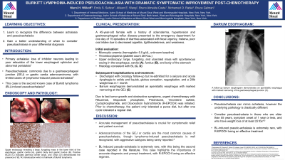
Case Description/Methods: A 45-year-old African American female with a history of scleroderma, hypertension and gastroesophageal reflux disease presented to the Emergency Department for 15 episodes of diarrhea associated with fecal urgency, melena, poor oral intake, lightheadedness and weakness. She denied weight loss or bleeding from other sites. Initial labs were notable for microcytic anemia (hemoglobin 5.5 g/dL from an unknown baseline) and thrombocytopenia (platelet count 28 K/uL). Upper endoscopy revealed a large, fungating and ulcerated mass with spontaneous oozing in the esophagus, cardia, fundus and body of the stomach. Histology was consistent with BL (CD20, CD10, MYC and BCL6 positive; Ki67 index 95%).
Upon discharge, the patient was scheduled to follow up with oncology, but was readmitted for a seizure. She was discharged and re-hospitalized for 4 days of vomiting and acute dysphagia to both solids and liquids. She had associated substernal globus sensation, food regurgitation within 2 minutes of consumption and a 28lb unintentional weight loss in 1 month. Barium esophagram demonstrated an aperistaltic esophagus with marked narrowing at the GEJ mimicking achalasia. Given the rapidly progressive and obstructive nature of the tumor, chemotherapy with rituximab, etoposide, prednisone, vincristine, cyclophosphamide and doxorubicin (R-EPOCH) was promptly initiated. Prior to chemotherapy, the patient only tolerated a pureed diet, but upon completion of one cycle, tolerated a regular diet.
Discussion: Accurate management of pseudoachalasia is crucial for symptomatic relief and patient survival. While adenocarcinomas of the GEJ or cardia are the most common cause, lymphoma-induced pseudoachalasia is well-recognized. Low-grade lymphomas account for most cases, and aggressive subtypes have been rarely reported. This is the second case of BL-induced pseudoachalasia reported in the literature and highlights the importance of accurate diagnosis and prompt treatment, with R-EPOCH being an effective regimen for inducing rapid responses.

Disclosures:
Marni Wilkoff, DO1, Emily S. Seltzer, DO, MS2, Allison E. Wang, MD3, Bruno A.. Costa, MD4, Mohamed O.. Rabie, MD5, Bruce Gelman, MD6. P3321 - Burkitt Lymphoma Induced Pseudoachalasia with Dramatic Symptomatic Improvement Post-Chemotherapy, ACG 2023 Annual Scientific Meeting Abstracts. Vancouver, BC, Canada: American College of Gastroenterology.
1Mount Sinai Morningside and West, New York, NY; 2Mount Sinai West and Morningside, New York, NY; 3Mount Sinai Morningside West, New York, NY; 4Mount Sinai Morningside and West, Icahn School of Medicine at Mount Sinai, New York, NY; 5Mount Sinai, New York, NY; 6Mount Sinai Morningside/West, New York, NY
Introduction: Primary achalasia is due to loss of inhibitory neurons leading to poor relaxation of the lower esophageal sphincter and abnormal peristalsis. In pseudoachalasia, a similar radiographic pattern derives from a secondary etiology, most commonly a gastroesophageal junction (GEJ) adenocarcinoma, though cases of lymphoma-induced pseudoachalasia have been reported. Here we present the second known case of Burkitt lymphoma (BL)-induced pseudoachalasia.
Case Description/Methods: A 45-year-old African American female with a history of scleroderma, hypertension and gastroesophageal reflux disease presented to the Emergency Department for 15 episodes of diarrhea associated with fecal urgency, melena, poor oral intake, lightheadedness and weakness. She denied weight loss or bleeding from other sites. Initial labs were notable for microcytic anemia (hemoglobin 5.5 g/dL from an unknown baseline) and thrombocytopenia (platelet count 28 K/uL). Upper endoscopy revealed a large, fungating and ulcerated mass with spontaneous oozing in the esophagus, cardia, fundus and body of the stomach. Histology was consistent with BL (CD20, CD10, MYC and BCL6 positive; Ki67 index 95%).
Upon discharge, the patient was scheduled to follow up with oncology, but was readmitted for a seizure. She was discharged and re-hospitalized for 4 days of vomiting and acute dysphagia to both solids and liquids. She had associated substernal globus sensation, food regurgitation within 2 minutes of consumption and a 28lb unintentional weight loss in 1 month. Barium esophagram demonstrated an aperistaltic esophagus with marked narrowing at the GEJ mimicking achalasia. Given the rapidly progressive and obstructive nature of the tumor, chemotherapy with rituximab, etoposide, prednisone, vincristine, cyclophosphamide and doxorubicin (R-EPOCH) was promptly initiated. Prior to chemotherapy, the patient only tolerated a pureed diet, but upon completion of one cycle, tolerated a regular diet.
Discussion: Accurate management of pseudoachalasia is crucial for symptomatic relief and patient survival. While adenocarcinomas of the GEJ or cardia are the most common cause, lymphoma-induced pseudoachalasia is well-recognized. Low-grade lymphomas account for most cases, and aggressive subtypes have been rarely reported. This is the second case of BL-induced pseudoachalasia reported in the literature and highlights the importance of accurate diagnosis and prompt treatment, with R-EPOCH being an effective regimen for inducing rapid responses.

Figure: Figure 1: Upper endoscopy revealing a large, fungating mass in the gastric cardia (A) and gastric fundus (B). Positive BCL6 (C) indicates germinal center origin, and c-Myc (D) demonstrates the presence of t(8;14) translocation which is hallmark of Burkitt Lymphoma. Barium esophagram demonstrates an aperistaltic esophagus with marked narrowing of the gastroesophageal junction (E).
Disclosures:
Marni Wilkoff indicated no relevant financial relationships.
Emily Seltzer indicated no relevant financial relationships.
Allison Wang indicated no relevant financial relationships.
Bruno Costa indicated no relevant financial relationships.
Mohamed Rabie indicated no relevant financial relationships.
Bruce Gelman indicated no relevant financial relationships.
Marni Wilkoff, DO1, Emily S. Seltzer, DO, MS2, Allison E. Wang, MD3, Bruno A.. Costa, MD4, Mohamed O.. Rabie, MD5, Bruce Gelman, MD6. P3321 - Burkitt Lymphoma Induced Pseudoachalasia with Dramatic Symptomatic Improvement Post-Chemotherapy, ACG 2023 Annual Scientific Meeting Abstracts. Vancouver, BC, Canada: American College of Gastroenterology.

