Tuesday Poster Session
Category: Esophagus
P3322 - A Rare Case of Esophageal Duplication Cyst With Esophago-Pleural Fistula and Pyopneumothorax
Tuesday, October 24, 2023
10:30 AM - 4:00 PM PT
Location: Exhibit Hall

Has Audio
- AA
Abdul Arham, MBBS, MD
UMass Chan-Baystate Medical Center
Springfield, MA
Presenting Author(s)
Award: Presidential Poster Award
Abdul Arham, MBBS, MD1, Sabika Iftikhar, MBBS, FCPS2, Saad Ur Rehman, BSc, MBBS2, Musfira Khalid, MBBS3, Faheem Javad, MBBS4, Abira Khalid, MBBS5, Abdul Waheed, MBBS6
1UMass Chan-Baystate Medical Center, Springfield, MA; 2Hameed Latif Hospital, Lahore, Punjab, Pakistan; 3Fatima Jinnah Medical University, Mississauga, ON, Canada; 4Al-Nafees Medical College, Mississauga, ON, Canada; 5Fatima Jinnah Medical University, Lahore, Punjab, Pakistan; 6Ameer-ud-Din Medical College, PGMI, Lahore, Punjab, Pakistan
Introduction: There are very few cases of congenital cervical esophageal duplication cysts, and even rare is esophagopleural fistula and pyopneumothorax associated with such cysts. Patient`s with cysts usually present with nonspecific symptoms like feeding difficulties, stridor, cough and respiratory distress. Our report describes what we believe may be a novel case from Pakistan of a 2-day-old boy whose respiratory distress gradually increased from birth onward.
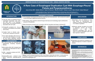
Case Description/Methods: The baby was delivered at term via spontaneous vaginal delivery with the weight of 3kg and APGARS 7/10 and 8/10 at 1 minutes and 5 minutes respectively. On day 2, he presented with respiratory difficulty - CXR showed right lower zone opacity and labs showed pH= 7.13, pCo2=55, pO2=80, Bicarbonate=15.8, and TLC=19.5. The patient was intubated, but on day 4 he developed a pneumothorax requiring chest tube placement resulting in an exudative mucoid discharge suggesting pyopneumothorax. There was gradual improvement with antibiotics and feed via nasogastric tube was started on 8th day of admission. However, with feeding, milky secretions drained through chest tube and microbiology report confirmed the presence of fat globules. Gastrograffin study showed contrast in lung pleura confirming the diagnosis of esophagopleural fistula. Neck thoracotomy was performed and an esophageal duplication cyst of 5 × 4 cm in anterior mediastinum was visualized. Following surgery his symptoms started improving, feeding resumed gradually, and he was discharged from hospital.
Discussion: Duplication cysts are believed to result from failure of vascularization and budding of primitive esophagus between 4 to 8 weeks of gestation (1) They are mostly found in thoracic region or below and rarely present as cervical. In Palmer's view, histology provides the best diagnostic tool in the form of three main criteria: attachment to esophageal wall, lining of GI mucosa, and presence of muscle layer. (10) Small cysts can go unnoticed until adulthood and may manifest as hematemesis, infection, difficulty swallowing, and rarely with malignant transformations or acute rupture of the cyst. (4,5,6) However, we report a very rare presentation of the cyst leading to fistula and pyopnemothorax and advise that duplication cysts be considered in the differential diagnosis of newborn respiratory distress.

Disclosures:
Abdul Arham, MBBS, MD1, Sabika Iftikhar, MBBS, FCPS2, Saad Ur Rehman, BSc, MBBS2, Musfira Khalid, MBBS3, Faheem Javad, MBBS4, Abira Khalid, MBBS5, Abdul Waheed, MBBS6. P3322 - A Rare Case of Esophageal Duplication Cyst With Esophago-Pleural Fistula and Pyopneumothorax, ACG 2023 Annual Scientific Meeting Abstracts. Vancouver, BC, Canada: American College of Gastroenterology.
Abdul Arham, MBBS, MD1, Sabika Iftikhar, MBBS, FCPS2, Saad Ur Rehman, BSc, MBBS2, Musfira Khalid, MBBS3, Faheem Javad, MBBS4, Abira Khalid, MBBS5, Abdul Waheed, MBBS6
1UMass Chan-Baystate Medical Center, Springfield, MA; 2Hameed Latif Hospital, Lahore, Punjab, Pakistan; 3Fatima Jinnah Medical University, Mississauga, ON, Canada; 4Al-Nafees Medical College, Mississauga, ON, Canada; 5Fatima Jinnah Medical University, Lahore, Punjab, Pakistan; 6Ameer-ud-Din Medical College, PGMI, Lahore, Punjab, Pakistan
Introduction: There are very few cases of congenital cervical esophageal duplication cysts, and even rare is esophagopleural fistula and pyopneumothorax associated with such cysts. Patient`s with cysts usually present with nonspecific symptoms like feeding difficulties, stridor, cough and respiratory distress. Our report describes what we believe may be a novel case from Pakistan of a 2-day-old boy whose respiratory distress gradually increased from birth onward.
Case Description/Methods: The baby was delivered at term via spontaneous vaginal delivery with the weight of 3kg and APGARS 7/10 and 8/10 at 1 minutes and 5 minutes respectively. On day 2, he presented with respiratory difficulty - CXR showed right lower zone opacity and labs showed pH= 7.13, pCo2=55, pO2=80, Bicarbonate=15.8, and TLC=19.5. The patient was intubated, but on day 4 he developed a pneumothorax requiring chest tube placement resulting in an exudative mucoid discharge suggesting pyopneumothorax. There was gradual improvement with antibiotics and feed via nasogastric tube was started on 8th day of admission. However, with feeding, milky secretions drained through chest tube and microbiology report confirmed the presence of fat globules. Gastrograffin study showed contrast in lung pleura confirming the diagnosis of esophagopleural fistula. Neck thoracotomy was performed and an esophageal duplication cyst of 5 × 4 cm in anterior mediastinum was visualized. Following surgery his symptoms started improving, feeding resumed gradually, and he was discharged from hospital.
Discussion: Duplication cysts are believed to result from failure of vascularization and budding of primitive esophagus between 4 to 8 weeks of gestation (1) They are mostly found in thoracic region or below and rarely present as cervical. In Palmer's view, histology provides the best diagnostic tool in the form of three main criteria: attachment to esophageal wall, lining of GI mucosa, and presence of muscle layer. (10) Small cysts can go unnoticed until adulthood and may manifest as hematemesis, infection, difficulty swallowing, and rarely with malignant transformations or acute rupture of the cyst. (4,5,6) However, we report a very rare presentation of the cyst leading to fistula and pyopnemothorax and advise that duplication cysts be considered in the differential diagnosis of newborn respiratory distress.

Figure: Figures: A and B showing Presence of milk in the tubing of chest tube and drain. C shows presence of contrast in lung and in tubing of chest tube through the duplication cyst. In D, arrow pointing towards the esophageal cyst.
Disclosures:
Abdul Arham indicated no relevant financial relationships.
Sabika Iftikhar indicated no relevant financial relationships.
Saad Ur Rehman indicated no relevant financial relationships.
Musfira Khalid indicated no relevant financial relationships.
Faheem Javad indicated no relevant financial relationships.
Abira Khalid indicated no relevant financial relationships.
Abdul Waheed indicated no relevant financial relationships.
Abdul Arham, MBBS, MD1, Sabika Iftikhar, MBBS, FCPS2, Saad Ur Rehman, BSc, MBBS2, Musfira Khalid, MBBS3, Faheem Javad, MBBS4, Abira Khalid, MBBS5, Abdul Waheed, MBBS6. P3322 - A Rare Case of Esophageal Duplication Cyst With Esophago-Pleural Fistula and Pyopneumothorax, ACG 2023 Annual Scientific Meeting Abstracts. Vancouver, BC, Canada: American College of Gastroenterology.

