Tuesday Poster Session
Category: Esophagus
P3323 - Esophageal Bullous Pemphigoid Presenting as Odynophagia
Tuesday, October 24, 2023
10:30 AM - 4:00 PM PT
Location: Exhibit Hall

Has Audio
- JL
Jessica Lin, MD, MBA
Johns Hopkins Hospital
Baltimore, MD
Presenting Author(s)
Jessica Lin, MD, MBA1, Danse Bi, MD1, Margaret G. Keane, MBBS, MSc2, Kathy Bull-Henry, MD1
1Johns Hopkins Hospital, Baltimore, MD; 2Johns Hopkins, Washington, DC
Introduction: Bullous pemphigoid is an autoimmune blistering disease that can affect the skin and mucous membranes. Mucosal involvement of the esophagus is rare and only a few cases have been reported in literature. Clinically, this can present as gastrointestinal bleeding, odynophagia, or dysphagia.
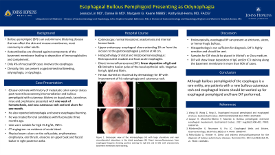
Case Description/Methods: An 83 year old male with a past medical history of metastatic adenocarcinoma of the colon status post recent ileocecetomy and chemoradiation and venous thromboembolism on apixaban presented with 1 week of hematochezia, new cutaneous rash and oral ulcers for one month. He had a known history of biopsy-confirmed bullous pemphigoid involving cutaneous blisters and was on dupulumab, tacrolimus rinse and prednisone. He subsequently developed odynophagia and had an additional episode of hematochezia on day 2 of hospitalization. Of note, he was also treated for oral candidiasis three months ago. Labs were notable for Hgb 15.8 g/dL and INR 1. CT angiogram showed no acute bleeding. On physical exam, he had an ulcer on the soft palate, erythematous oropharynx, oral thrush, erosions on upper back and flaccid bullae in the right posterior axilla. He had no ocular symptoms. He was started on fluconazole for presumed esophageal candidiasis but had no improvement after 72 hours. Colonoscopy showed normal ileocolonic anastomosis and internal hemorrhoids. Upper endoscopy showed esophageal ulcers extending 30 cm from the incisors to the gastroesophageal junction at 40 cm. Biopsies of the distal and mid/proximal esophagus showed fibrinopurulent exudate and focal acute esophagitis. Direct immunofluorescence (DIF) showed linear deposition of IgG and C3 limited to basilar poles of the basal epithelial cells which is pathognomic for bullous pemphigoid. He was started on rituximab by dermatology for bullous pemphigoid.
Discussion: Bullous pemphigoid is an acquired autoimmune disease that commonly affect patients older than 60 years old. Mucosal involvement is not common and only 4% of mucosal cases involves the esophagus. Endoscopically, esophageal bullous pemphigoid can present as strictures, ulcers, or hemorrhagic bullous. Although bullous pemphigoid of the esophagus is a rare entity, any patients with a new bullous cutaneous rash and esophageal lesions should be worked up for esophageal pemphigoid and DIF performed. It is important that samples for DIF are placed in Michel’s or Zeus medium in order to make the correct diagnosis.

Disclosures:
Jessica Lin, MD, MBA1, Danse Bi, MD1, Margaret G. Keane, MBBS, MSc2, Kathy Bull-Henry, MD1. P3323 - Esophageal Bullous Pemphigoid Presenting as Odynophagia, ACG 2023 Annual Scientific Meeting Abstracts. Vancouver, BC, Canada: American College of Gastroenterology.
1Johns Hopkins Hospital, Baltimore, MD; 2Johns Hopkins, Washington, DC
Introduction: Bullous pemphigoid is an autoimmune blistering disease that can affect the skin and mucous membranes. Mucosal involvement of the esophagus is rare and only a few cases have been reported in literature. Clinically, this can present as gastrointestinal bleeding, odynophagia, or dysphagia.
Case Description/Methods: An 83 year old male with a past medical history of metastatic adenocarcinoma of the colon status post recent ileocecetomy and chemoradiation and venous thromboembolism on apixaban presented with 1 week of hematochezia, new cutaneous rash and oral ulcers for one month. He had a known history of biopsy-confirmed bullous pemphigoid involving cutaneous blisters and was on dupulumab, tacrolimus rinse and prednisone. He subsequently developed odynophagia and had an additional episode of hematochezia on day 2 of hospitalization. Of note, he was also treated for oral candidiasis three months ago. Labs were notable for Hgb 15.8 g/dL and INR 1. CT angiogram showed no acute bleeding. On physical exam, he had an ulcer on the soft palate, erythematous oropharynx, oral thrush, erosions on upper back and flaccid bullae in the right posterior axilla. He had no ocular symptoms. He was started on fluconazole for presumed esophageal candidiasis but had no improvement after 72 hours. Colonoscopy showed normal ileocolonic anastomosis and internal hemorrhoids. Upper endoscopy showed esophageal ulcers extending 30 cm from the incisors to the gastroesophageal junction at 40 cm. Biopsies of the distal and mid/proximal esophagus showed fibrinopurulent exudate and focal acute esophagitis. Direct immunofluorescence (DIF) showed linear deposition of IgG and C3 limited to basilar poles of the basal epithelial cells which is pathognomic for bullous pemphigoid. He was started on rituximab by dermatology for bullous pemphigoid.
Discussion: Bullous pemphigoid is an acquired autoimmune disease that commonly affect patients older than 60 years old. Mucosal involvement is not common and only 4% of mucosal cases involves the esophagus. Endoscopically, esophageal bullous pemphigoid can present as strictures, ulcers, or hemorrhagic bullous. Although bullous pemphigoid of the esophagus is a rare entity, any patients with a new bullous cutaneous rash and esophageal lesions should be worked up for esophageal pemphigoid and DIF performed. It is important that samples for DIF are placed in Michel’s or Zeus medium in order to make the correct diagnosis.

Figure: Figure 1: Endoscopic view of the mid-esophagus (a) with large ulcerations and near circumferential ulcerations in the distal esophagus (b)
Disclosures:
Jessica Lin indicated no relevant financial relationships.
Danse Bi indicated no relevant financial relationships.
Margaret Keane indicated no relevant financial relationships.
Kathy Bull-Henry indicated no relevant financial relationships.
Jessica Lin, MD, MBA1, Danse Bi, MD1, Margaret G. Keane, MBBS, MSc2, Kathy Bull-Henry, MD1. P3323 - Esophageal Bullous Pemphigoid Presenting as Odynophagia, ACG 2023 Annual Scientific Meeting Abstracts. Vancouver, BC, Canada: American College of Gastroenterology.
