Tuesday Poster Session
Category: Esophagus
P3329 - Novelty in North American: Small Cell Esophageal Cancer in the Background of Barrett’s Esophagus
Tuesday, October 24, 2023
10:30 AM - 4:00 PM PT
Location: Exhibit Hall

Has Audio

Rohan Rau, DO
Albert Einstein Internal Medicine Residency
Philadelphia, PA
Presenting Author(s)
Rohan Rau, DO1, Julie Sullivan, DO2, Michael Goldberg, DO3, Sanjay Rau, DO4
1Albert Einstein Internal Medicine Residency, Philadelphia, PA; 2Albert Einstein Gastroenterology Fellowship, Philadelphia, PA; 3Albert Einstein Gastroenterology, Philadelphia, PA; 4Jefferson Health, Philadelphia, PA
Introduction: Barrett’s esophagus (BE) is a state of dysplasia as esophageal squamous epithelium transforms into columnar epithelium. These precancerous lesions can progress into adenocarcinoma, which is the predominant esophageal malignancy in the United States. In contrast, small cell esophageal carcinoma (SCEC) accounts for less than 1% of esophageal malignancies. An even rarer event is SCEC developing in the background of BE. We present the first case in North America of a patient with Barrett’s esophagus who developed primary small cell esophageal carcinoma.
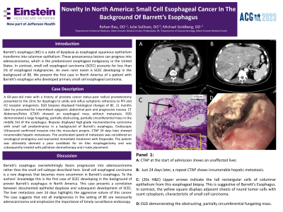
Case Description/Methods: A 60-year-old male with a history of prostate cancer status-post radical prostatectomy presented to the clinic for dysphagia to solids and reflux symptoms refractory to PPI and H2 receptor antagonists. EGD biopsies displayed the characteristic histological changes of BE. 11 months later he presented for intermittent epigastric abdominal pain and progressive nausea. CT Abdomen/Pelvis (CTAP) showed an esophageal mass without metastasis. EGD demonstrated a large fungating, partially obstructing, partially circumferential mass in the middle 3rd of the esophagus. Biopsies displayed high-grade neuroendocrine carcinoma with small cell predominance in a background of Barrett's esophagus. Endoscopic Ultrasound confirmed invasion into the muscularis propria. CTAP 24 days later showed innumerable hepatic metastases. The accelerated speed of metastasis was considered an oncological emergency and warranted immediate treatment with Etoposide. The patient was ultimately deemed a poor candidate for en bloc esophagectomy and was subsequently treated with palliative chemotherapy and J tube placement.
Discussion: Barrett's esophagus overwhelmingly favors progression into adenocarcinoma rather than the small cell subtype described here. Small cell esophageal carcinoma is a rare diagnosis that becomes more uncommon in Barrett's esophagus. To the authors’ knowledge this is the first reported case of SCEC developing in the background of proven Barrett’s esophagus in North America. This case presents a correlation between documented epithelial dysplasia and subsequent development of SCEC. The liver metastasis over 24 days highlights the aggressive nature of this cancer. The case suggests that not all malignancies in the setting of BE are necessarily adenocarcinoma and emphasizes the importance of timely surveillance endoscopy.

Disclosures:
Rohan Rau, DO1, Julie Sullivan, DO2, Michael Goldberg, DO3, Sanjay Rau, DO4. P3329 - Novelty in North American: Small Cell Esophageal Cancer in the Background of Barrett’s Esophagus, ACG 2023 Annual Scientific Meeting Abstracts. Vancouver, BC, Canada: American College of Gastroenterology.
1Albert Einstein Internal Medicine Residency, Philadelphia, PA; 2Albert Einstein Gastroenterology Fellowship, Philadelphia, PA; 3Albert Einstein Gastroenterology, Philadelphia, PA; 4Jefferson Health, Philadelphia, PA
Introduction: Barrett’s esophagus (BE) is a state of dysplasia as esophageal squamous epithelium transforms into columnar epithelium. These precancerous lesions can progress into adenocarcinoma, which is the predominant esophageal malignancy in the United States. In contrast, small cell esophageal carcinoma (SCEC) accounts for less than 1% of esophageal malignancies. An even rarer event is SCEC developing in the background of BE. We present the first case in North America of a patient with Barrett’s esophagus who developed primary small cell esophageal carcinoma.
Case Description/Methods: A 60-year-old male with a history of prostate cancer status-post radical prostatectomy presented to the clinic for dysphagia to solids and reflux symptoms refractory to PPI and H2 receptor antagonists. EGD biopsies displayed the characteristic histological changes of BE. 11 months later he presented for intermittent epigastric abdominal pain and progressive nausea. CT Abdomen/Pelvis (CTAP) showed an esophageal mass without metastasis. EGD demonstrated a large fungating, partially obstructing, partially circumferential mass in the middle 3rd of the esophagus. Biopsies displayed high-grade neuroendocrine carcinoma with small cell predominance in a background of Barrett's esophagus. Endoscopic Ultrasound confirmed invasion into the muscularis propria. CTAP 24 days later showed innumerable hepatic metastases. The accelerated speed of metastasis was considered an oncological emergency and warranted immediate treatment with Etoposide. The patient was ultimately deemed a poor candidate for en bloc esophagectomy and was subsequently treated with palliative chemotherapy and J tube placement.
Discussion: Barrett's esophagus overwhelmingly favors progression into adenocarcinoma rather than the small cell subtype described here. Small cell esophageal carcinoma is a rare diagnosis that becomes more uncommon in Barrett's esophagus. To the authors’ knowledge this is the first reported case of SCEC developing in the background of proven Barrett’s esophagus in North America. This case presents a correlation between documented epithelial dysplasia and subsequent development of SCEC. The liver metastasis over 24 days highlights the aggressive nature of this cancer. The case suggests that not all malignancies in the setting of BE are necessarily adenocarcinoma and emphasizes the importance of timely surveillance endoscopy.

Figure: A: CTAP at start of admission shows an unaffected liver. B: By day 24, repeat CTAP shows innumerable hepatic metastasis. C: (20x H&E) Upper arrows indicate the tall rectangular cells of columnar epithelium from this esophageal biopsy. This is suggestive of Barrett’s Esophagus. In contrast, the yellow square displays adjacent sheets of round tumor cells with scant cytoplasm, characteristic of small cell carcinoma. D: EGD demonstrating the obstructing, partially circumferential fungating mass.
Disclosures:
Rohan Rau indicated no relevant financial relationships.
Julie Sullivan indicated no relevant financial relationships.
Michael Goldberg indicated no relevant financial relationships.
Sanjay Rau indicated no relevant financial relationships.
Rohan Rau, DO1, Julie Sullivan, DO2, Michael Goldberg, DO3, Sanjay Rau, DO4. P3329 - Novelty in North American: Small Cell Esophageal Cancer in the Background of Barrett’s Esophagus, ACG 2023 Annual Scientific Meeting Abstracts. Vancouver, BC, Canada: American College of Gastroenterology.

