Tuesday Poster Session
Category: Esophagus
P3333 - Tearing the Norm: Mallory-Weiss Unveiled - Dysphagia as a Surprising Presentation
Tuesday, October 24, 2023
10:30 AM - 4:00 PM PT
Location: Exhibit Hall

Has Audio

Mohammad Nabil Rayad, MD
Saint Michael's Medical Center, New York Medical College
Newark, New Jersey
Presenting Author(s)
Mohammad Nabil Rayad, MD1, Noreen Mirza, MD1, Vikramjit S. Purewal, DO2, Dema Shamoon, MD1, Muhammad Hussain, MD1, Yatinder Bains, MD1
1Saint Michael's Medical Center, New York Medical College, Newark, NJ; 2Mountain Vista Medical Center, Mesa, AZ
Introduction: Mallory-Weiss tear (MWT) is a a tear in the lining of the gastroesophageal junction caused by excessive pressure on the area due to retching, coughing or vomiting. It can account for around 5-15% of upper gastrointestinal bleeding cases. Herein, we present a rare case of MWT presenting as dysphagia due to massive blood clot.
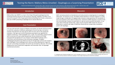
Case Description/Methods: A 47 year-old-male with a history of diabetes and alcohol use disorder (AUD) presented to the ED for evaluation of difficulty swallowing for the last two days. He had one episode of hematemesis two days ago for which he never went to the hospital for evaluation. Vital signs were normal and physical examination was significant for epigastric tenderness. Initial hemoglobin was 7.3 g/dL. The electrolytes, kidney function were normal and LFTs were mildly elevated with normal bilirubin and PT/INR. Computerized tomography (CT) scan showed a mildly dilated esophagus with a 3.6x2.3x5cm soft tissue density in the mid thoracic esophagus. Emergent esophagogastroduodenoscopy (EGD) was done for foreign body removal and revealed a web, esophagitis, three esophageal ulcers with two actively bleeding and a large clot was seen surrounding one proximal ulcer which was removed by cold snare. The actively bleeding ulcers were treated with coagulation and hemostatic clips. His dysphagia resolved post-procedure.
Discussion: MWT commonly presents as hematemesis and rarely presents as dysphagia due to esophageal obstruction. If it presents as such, immediate medical attention is required. Diagnosis is usually made through a combination of imaging studies; however, endoscopy allows for visualization of tears and evaluation of the extent of obstruction. Treatment of MWT depends on severity of the tear and endoscopic therapy via cauterization or clips are used to promote hemostasis. In rare cases where endoscopic therapy fails, surgery may be required. MWT should not only be a differential in a patient with upper GI bleed but should also be included when evaluating a patient with dysphagia.

Disclosures:
Mohammad Nabil Rayad, MD1, Noreen Mirza, MD1, Vikramjit S. Purewal, DO2, Dema Shamoon, MD1, Muhammad Hussain, MD1, Yatinder Bains, MD1. P3333 - Tearing the Norm: Mallory-Weiss Unveiled - Dysphagia as a Surprising Presentation, ACG 2023 Annual Scientific Meeting Abstracts. Vancouver, BC, Canada: American College of Gastroenterology.
1Saint Michael's Medical Center, New York Medical College, Newark, NJ; 2Mountain Vista Medical Center, Mesa, AZ
Introduction: Mallory-Weiss tear (MWT) is a a tear in the lining of the gastroesophageal junction caused by excessive pressure on the area due to retching, coughing or vomiting. It can account for around 5-15% of upper gastrointestinal bleeding cases. Herein, we present a rare case of MWT presenting as dysphagia due to massive blood clot.
Case Description/Methods: A 47 year-old-male with a history of diabetes and alcohol use disorder (AUD) presented to the ED for evaluation of difficulty swallowing for the last two days. He had one episode of hematemesis two days ago for which he never went to the hospital for evaluation. Vital signs were normal and physical examination was significant for epigastric tenderness. Initial hemoglobin was 7.3 g/dL. The electrolytes, kidney function were normal and LFTs were mildly elevated with normal bilirubin and PT/INR. Computerized tomography (CT) scan showed a mildly dilated esophagus with a 3.6x2.3x5cm soft tissue density in the mid thoracic esophagus. Emergent esophagogastroduodenoscopy (EGD) was done for foreign body removal and revealed a web, esophagitis, three esophageal ulcers with two actively bleeding and a large clot was seen surrounding one proximal ulcer which was removed by cold snare. The actively bleeding ulcers were treated with coagulation and hemostatic clips. His dysphagia resolved post-procedure.
Discussion: MWT commonly presents as hematemesis and rarely presents as dysphagia due to esophageal obstruction. If it presents as such, immediate medical attention is required. Diagnosis is usually made through a combination of imaging studies; however, endoscopy allows for visualization of tears and evaluation of the extent of obstruction. Treatment of MWT depends on severity of the tear and endoscopic therapy via cauterization or clips are used to promote hemostasis. In rare cases where endoscopic therapy fails, surgery may be required. MWT should not only be a differential in a patient with upper GI bleed but should also be included when evaluating a patient with dysphagia.

Figure: A = Esophageal web
B= Bleeding esophageal ulcer
C and D= Esophagitis with ulcers
E= CT chest showing mid dilated esophagus with presence of 3.6 x2.3 x5cm soft tissue density in mid thoracic esophagus
F= Gross image of large blood clot removed from esophagus
B= Bleeding esophageal ulcer
C and D= Esophagitis with ulcers
E= CT chest showing mid dilated esophagus with presence of 3.6 x2.3 x5cm soft tissue density in mid thoracic esophagus
F= Gross image of large blood clot removed from esophagus
Disclosures:
Mohammad Nabil Rayad indicated no relevant financial relationships.
Noreen Mirza indicated no relevant financial relationships.
Vikramjit Purewal indicated no relevant financial relationships.
Dema Shamoon indicated no relevant financial relationships.
Muhammad Hussain indicated no relevant financial relationships.
Yatinder Bains indicated no relevant financial relationships.
Mohammad Nabil Rayad, MD1, Noreen Mirza, MD1, Vikramjit S. Purewal, DO2, Dema Shamoon, MD1, Muhammad Hussain, MD1, Yatinder Bains, MD1. P3333 - Tearing the Norm: Mallory-Weiss Unveiled - Dysphagia as a Surprising Presentation, ACG 2023 Annual Scientific Meeting Abstracts. Vancouver, BC, Canada: American College of Gastroenterology.
