Tuesday Poster Session
Category: Esophagus
P3339 - Reliance on Clinical Judgment to Target Immunohistochemical Stain Use for Diagnosis of HSV Esophagitis
Tuesday, October 24, 2023
10:30 AM - 4:00 PM PT
Location: Exhibit Hall

Has Audio

Aastha V. Bharwad, MD
University of Kansas School of Medicine
Wichita, KS
Presenting Author(s)
Aastha Bharwad, MD, Lawrence Zhou, MD, William J.. Salyers, MD, MPH
University of Kansas School of Medicine, Wichita, KS
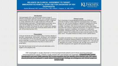
Introduction: Viral esophagitis (VE) is the second most common cause of infectious esophagitis, following Candida, and is primarily caused by herpes simplex virus (HSV) and cytomegalovirus (CMV). Diagnosis is usually established with an upper endoscopy confirmed with histopathological examination. Histopathological examination alone may miss the diagnosis and tissue viral culture enhances the diagnostic sensitivity. We discuss the case of a patient with negative histology and culture results, who eventually tested positive for HSV with immunohistochemistry (IHC).
Case Description/Methods: A 39-year-old female with a past medical history of anxiety, allergies to penicillin, sulfacetamide, nitrofurantoin, levofloxacin, and undergoing in vitro fertilization (IVF) for the past year presented to our clinic for severe dysphagia. As part of her IVF, the patient had completed a 10-day course of dexamethasone 2mg daily six weeks prior to presentation. Initial lab results revealed normal cell counts and electrolytes, and a negative urine pregnancy test. Due to dysphagia, an esophagogastroduodenoscopy (EGD) was performed revealing multiple superficial esophageal ulcers, the largest being 8 mm, with no active bleeding or stigmata of bleeding and normal duodenum. The patient was started on a proton pump inhibitor and sucralfate. The biopsy of the ulcers showed severe active inflammation associated with numerous segmented neutrophils, but negative for Barrett’s esophagus, neoplasm or dysplasia, viral inclusions, or eosinophilic esophagitis. Subsequent fungal, CMV, and HSV culture of the ulcer was negative, however immunohistochemical stains returned positive for HSV types I and II. The patient denied any history of oral or genital lesions.
The patient was started on a 10-day course of valacyclovir with eventual resolution of symptoms following treatment. Subsequent workup to rule out other causes of immunosuppression showed negative HIV-1 viral load and 4th generation HIV screen, normal IgG, IgA, and IgM, and normal C3, C4, and CH50 complement and negative anti-neutrophil antibody.
Discussion: HSV esophagitis is usually observed in immunocompromised patients and in those receiving prolonged immunosuppressive agents. Patients usually present with odynophagia and dysphagia. Compared with routine hematoxylin and eosin (H&E) staining, IHC is more sensitive and can identify more infected cells. IHC stains should primarily be employed in cases where histopathologic findings are ambiguous and clinical suspicion remains high.
Disclosures:
Aastha Bharwad, MD, Lawrence Zhou, MD, William J.. Salyers, MD, MPH. P3339 - Reliance on Clinical Judgment to Target Immunohistochemical Stain Use for Diagnosis of HSV Esophagitis, ACG 2023 Annual Scientific Meeting Abstracts. Vancouver, BC, Canada: American College of Gastroenterology.
University of Kansas School of Medicine, Wichita, KS
Introduction: Viral esophagitis (VE) is the second most common cause of infectious esophagitis, following Candida, and is primarily caused by herpes simplex virus (HSV) and cytomegalovirus (CMV). Diagnosis is usually established with an upper endoscopy confirmed with histopathological examination. Histopathological examination alone may miss the diagnosis and tissue viral culture enhances the diagnostic sensitivity. We discuss the case of a patient with negative histology and culture results, who eventually tested positive for HSV with immunohistochemistry (IHC).
Case Description/Methods: A 39-year-old female with a past medical history of anxiety, allergies to penicillin, sulfacetamide, nitrofurantoin, levofloxacin, and undergoing in vitro fertilization (IVF) for the past year presented to our clinic for severe dysphagia. As part of her IVF, the patient had completed a 10-day course of dexamethasone 2mg daily six weeks prior to presentation. Initial lab results revealed normal cell counts and electrolytes, and a negative urine pregnancy test. Due to dysphagia, an esophagogastroduodenoscopy (EGD) was performed revealing multiple superficial esophageal ulcers, the largest being 8 mm, with no active bleeding or stigmata of bleeding and normal duodenum. The patient was started on a proton pump inhibitor and sucralfate. The biopsy of the ulcers showed severe active inflammation associated with numerous segmented neutrophils, but negative for Barrett’s esophagus, neoplasm or dysplasia, viral inclusions, or eosinophilic esophagitis. Subsequent fungal, CMV, and HSV culture of the ulcer was negative, however immunohistochemical stains returned positive for HSV types I and II. The patient denied any history of oral or genital lesions.
The patient was started on a 10-day course of valacyclovir with eventual resolution of symptoms following treatment. Subsequent workup to rule out other causes of immunosuppression showed negative HIV-1 viral load and 4th generation HIV screen, normal IgG, IgA, and IgM, and normal C3, C4, and CH50 complement and negative anti-neutrophil antibody.
Discussion: HSV esophagitis is usually observed in immunocompromised patients and in those receiving prolonged immunosuppressive agents. Patients usually present with odynophagia and dysphagia. Compared with routine hematoxylin and eosin (H&E) staining, IHC is more sensitive and can identify more infected cells. IHC stains should primarily be employed in cases where histopathologic findings are ambiguous and clinical suspicion remains high.
Disclosures:
Aastha Bharwad indicated no relevant financial relationships.
Lawrence Zhou indicated no relevant financial relationships.
William Salyers indicated no relevant financial relationships.
Aastha Bharwad, MD, Lawrence Zhou, MD, William J.. Salyers, MD, MPH. P3339 - Reliance on Clinical Judgment to Target Immunohistochemical Stain Use for Diagnosis of HSV Esophagitis, ACG 2023 Annual Scientific Meeting Abstracts. Vancouver, BC, Canada: American College of Gastroenterology.
