Tuesday Poster Session
Category: Esophagus
P3340 - Rare Case of Esophageal Ulcers Secondary to Thermal Injury After Radiofrequency Ablation for Atrial Fibrillation
Tuesday, October 24, 2023
10:30 AM - 4:00 PM PT
Location: Exhibit Hall

Has Audio

Aastha Chokshi, MD
Brown University
Providence, RI
Presenting Author(s)
Aastha Chokshi, MD1, Averill Guo, MD2, Fadlallah Habr, MD3
1Brown University, Providence, RI; 2Brown University/Warren Alpert Medical School, Providence, RI; 3Brown University/Lifespan Physician Group, East Providence, RI
Introduction: Catheter radiofrequency ablation is a commonly used procedure for the treatment of patients with atrial fibrillation (AF). The proximity of the esophagus to the left atrium increases the risk of esophageal injury, which can range from minor erythema to fatal esophageal perforation or atrioesophageal fistula formation. Such thermal injuries are often underdiagnosed and guidelines on prevention and surveillance are limited due to a lack of randomized controlled trials. While endoscopically detectable asymptomatic esophageal lesions may be identified in up to 20-40% of cases, ulcers are less common and have a 10% risk of progression to perforation. It is essential to identify at-risk patients early to prevent complications.
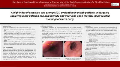
Case Description/Methods: We describe a case of a 62-year-old man with past medical history of hypertension, heart failure with reduced ejection fraction, and newly diagnosed atrial fibrillation undergoing a radiofrequency ablation. Throughout the procedure, an esophageal temperature probe was used to monitor for esophageal heating. Esophageal heating to a temperature of 39.5oC was noted during isolation of the left inferior pulmonary vein, and thus ablation adjacent to the esophagus was not performed. The patient underwent an esophagoduodenoscopy (EGD) which showed a nonbleeding esophageal ulcer 30 cm from the incisors at the level of the left atrium (Figure 1A). The patient was started on pantoprazole 40 mg twice daily, sucralfate 1 g four times daily, and counseled to maintain a liquid diet, avoiding coarse or potentially sharp foods. A repeat EGD showed a nonbleeding esophageal erosion in the same location, which appeared improved from 1 week prior (Figure 1B). The patient was continued on pantoprazole 40 mg twice daily for 8 weeks.
Discussion: Our case highlights that a high index of suspicion and prompt EGD evaluation in at risk patients undergoing radiofrequency ablation can help identify and intervene upon thermal injury related esophageal ulcers early. Risk factors for thermal esophageal injuries include elevated maximum esophageal temperature, anatomic variation, catheter tip size, contact pressure and catheter orientation among others. It is important to recognize risk factors in real time and to identify and closely monitor high risk patients, especially given the significant morbidity and mortality associated with perforating complications. Additional studies that aim to establish criteria for EGD evaluation are needed.

Disclosures:
Aastha Chokshi, MD1, Averill Guo, MD2, Fadlallah Habr, MD3. P3340 - Rare Case of Esophageal Ulcers Secondary to Thermal Injury After Radiofrequency Ablation for Atrial Fibrillation, ACG 2023 Annual Scientific Meeting Abstracts. Vancouver, BC, Canada: American College of Gastroenterology.
1Brown University, Providence, RI; 2Brown University/Warren Alpert Medical School, Providence, RI; 3Brown University/Lifespan Physician Group, East Providence, RI
Introduction: Catheter radiofrequency ablation is a commonly used procedure for the treatment of patients with atrial fibrillation (AF). The proximity of the esophagus to the left atrium increases the risk of esophageal injury, which can range from minor erythema to fatal esophageal perforation or atrioesophageal fistula formation. Such thermal injuries are often underdiagnosed and guidelines on prevention and surveillance are limited due to a lack of randomized controlled trials. While endoscopically detectable asymptomatic esophageal lesions may be identified in up to 20-40% of cases, ulcers are less common and have a 10% risk of progression to perforation. It is essential to identify at-risk patients early to prevent complications.
Case Description/Methods: We describe a case of a 62-year-old man with past medical history of hypertension, heart failure with reduced ejection fraction, and newly diagnosed atrial fibrillation undergoing a radiofrequency ablation. Throughout the procedure, an esophageal temperature probe was used to monitor for esophageal heating. Esophageal heating to a temperature of 39.5oC was noted during isolation of the left inferior pulmonary vein, and thus ablation adjacent to the esophagus was not performed. The patient underwent an esophagoduodenoscopy (EGD) which showed a nonbleeding esophageal ulcer 30 cm from the incisors at the level of the left atrium (Figure 1A). The patient was started on pantoprazole 40 mg twice daily, sucralfate 1 g four times daily, and counseled to maintain a liquid diet, avoiding coarse or potentially sharp foods. A repeat EGD showed a nonbleeding esophageal erosion in the same location, which appeared improved from 1 week prior (Figure 1B). The patient was continued on pantoprazole 40 mg twice daily for 8 weeks.
Discussion: Our case highlights that a high index of suspicion and prompt EGD evaluation in at risk patients undergoing radiofrequency ablation can help identify and intervene upon thermal injury related esophageal ulcers early. Risk factors for thermal esophageal injuries include elevated maximum esophageal temperature, anatomic variation, catheter tip size, contact pressure and catheter orientation among others. It is important to recognize risk factors in real time and to identify and closely monitor high risk patients, especially given the significant morbidity and mortality associated with perforating complications. Additional studies that aim to establish criteria for EGD evaluation are needed.

Figure: Figure 1. A) Esophageal ulcer noted on initial EGD 30 cm from incisors at level of left atrium. B) Esophageal Ulcer noted on follow up EGD, found to be improved from prior.
Disclosures:
Aastha Chokshi indicated no relevant financial relationships.
Averill Guo indicated no relevant financial relationships.
Fadlallah Habr indicated no relevant financial relationships.
Aastha Chokshi, MD1, Averill Guo, MD2, Fadlallah Habr, MD3. P3340 - Rare Case of Esophageal Ulcers Secondary to Thermal Injury After Radiofrequency Ablation for Atrial Fibrillation, ACG 2023 Annual Scientific Meeting Abstracts. Vancouver, BC, Canada: American College of Gastroenterology.
