Tuesday Poster Session
Category: Esophagus
P3342 - Dilated Azygous Vein Secondary to Inferior Vena Cava Atresia as a Rare Cause of Dysphagia
Tuesday, October 24, 2023
10:30 AM - 4:00 PM PT
Location: Exhibit Hall

Has Audio
- RM
Rasheed Musa, MD
East Tennessee State University
Johnson City, TN
Presenting Author(s)
Award: Presidential Poster Award
Rasheed Musa, MD1, Asaiel Makahleh, MD2, Fouad Jaber, MD3, Lana Makahleh, MD1, Jason McKinney, DO4
1East Tennessee State University, Johnson City, TN; 2East Tennesse State University, Johnson City, TN; 3University of Missouri-Kansas City, Kansas City, MO; 4East Tennessee State University / GI Associates of Northeast Tennessee, Johnson City, TN
Introduction: Dysphagia secondary to vascular abnormalities is a rare condition, with a prevalence of less than 1%, that occurs through secondary compression of the esophagus posteriorly by an abnormal intrathoracic vascular structure. It commonly presents with dysphagia to solids. Management is usually done with dietary modification; however, more severe, and intractable cases may require surgical intervention. We describe this rare entity in a 56-year-old male presenting with mechanical dysphagia secondary to dilated azygous and hemi-azygous vein resulting from chronic IVC stenosis.
Case Description/Methods: A 56-year-old male with a long-standing history of recurrent dysphagia for solids along with other comorbidities including CAD with cardiomyopathy and atrial fibrillation on long-term anticoagulation presents with recurrent debilitating dysphagia associated with weight loss.
The patient’s initial evaluation included a barium esophagogram that showed and small size sliding hiatal hernia as well as an esophageal ring, which was followed by went through multiple endoscopic evaluations with an EGD and dilation that did not show evidence of an actual ring and resulted in very minimal improvement in symptoms most recently one week prior to presentation.
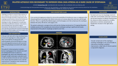
Cross-sectional imaging was ordered to role out the possibility of intrathoracic mass or malignancy that showed significant narrowing of the inferior vena cava as it courses above the renal vein and through the liver. There is a significantly dilated azygous and hemiazygous vein that compresses the esophagus between the aorta, left atrium, and azygous vein.
The patient underwent a venogram that confirmed the findings mentioned on CT imaging followed by multiple sessions of IVC venoplasty by interventional radiology resulting in significant and steady improvement of the patient’s dysphagia symptoms over time.
Discussion: Persistent and recurrent dysphagia mainly for solids with unremarkable endoscopic evaluation and minimal response to dilation should prompt the clinician to either evaluate for a motility dysfunction or extrinsic compression by intrathoracic structure in the right clinical setting.
Conclusion:
Vascular compression of the esophagus resulting in dysphagia is rare, yet amenable to intervention resulting in symptoms improvement, a high index of clinical suspicion id required for diagnosis.

Disclosures:
Rasheed Musa, MD1, Asaiel Makahleh, MD2, Fouad Jaber, MD3, Lana Makahleh, MD1, Jason McKinney, DO4. P3342 - Dilated Azygous Vein Secondary to Inferior Vena Cava Atresia as a Rare Cause of Dysphagia, ACG 2023 Annual Scientific Meeting Abstracts. Vancouver, BC, Canada: American College of Gastroenterology.
Rasheed Musa, MD1, Asaiel Makahleh, MD2, Fouad Jaber, MD3, Lana Makahleh, MD1, Jason McKinney, DO4
1East Tennessee State University, Johnson City, TN; 2East Tennesse State University, Johnson City, TN; 3University of Missouri-Kansas City, Kansas City, MO; 4East Tennessee State University / GI Associates of Northeast Tennessee, Johnson City, TN
Introduction: Dysphagia secondary to vascular abnormalities is a rare condition, with a prevalence of less than 1%, that occurs through secondary compression of the esophagus posteriorly by an abnormal intrathoracic vascular structure. It commonly presents with dysphagia to solids. Management is usually done with dietary modification; however, more severe, and intractable cases may require surgical intervention. We describe this rare entity in a 56-year-old male presenting with mechanical dysphagia secondary to dilated azygous and hemi-azygous vein resulting from chronic IVC stenosis.
Case Description/Methods: A 56-year-old male with a long-standing history of recurrent dysphagia for solids along with other comorbidities including CAD with cardiomyopathy and atrial fibrillation on long-term anticoagulation presents with recurrent debilitating dysphagia associated with weight loss.
The patient’s initial evaluation included a barium esophagogram that showed and small size sliding hiatal hernia as well as an esophageal ring, which was followed by went through multiple endoscopic evaluations with an EGD and dilation that did not show evidence of an actual ring and resulted in very minimal improvement in symptoms most recently one week prior to presentation.
Cross-sectional imaging was ordered to role out the possibility of intrathoracic mass or malignancy that showed significant narrowing of the inferior vena cava as it courses above the renal vein and through the liver. There is a significantly dilated azygous and hemiazygous vein that compresses the esophagus between the aorta, left atrium, and azygous vein.
The patient underwent a venogram that confirmed the findings mentioned on CT imaging followed by multiple sessions of IVC venoplasty by interventional radiology resulting in significant and steady improvement of the patient’s dysphagia symptoms over time.
Discussion: Persistent and recurrent dysphagia mainly for solids with unremarkable endoscopic evaluation and minimal response to dilation should prompt the clinician to either evaluate for a motility dysfunction or extrinsic compression by intrathoracic structure in the right clinical setting.
Conclusion:
Vascular compression of the esophagus resulting in dysphagia is rare, yet amenable to intervention resulting in symptoms improvement, a high index of clinical suspicion id required for diagnosis.

Figure: Cross-sectional imaging
Disclosures:
Rasheed Musa indicated no relevant financial relationships.
Asaiel Makahleh indicated no relevant financial relationships.
Fouad Jaber indicated no relevant financial relationships.
Lana Makahleh indicated no relevant financial relationships.
Jason McKinney indicated no relevant financial relationships.
Rasheed Musa, MD1, Asaiel Makahleh, MD2, Fouad Jaber, MD3, Lana Makahleh, MD1, Jason McKinney, DO4. P3342 - Dilated Azygous Vein Secondary to Inferior Vena Cava Atresia as a Rare Cause of Dysphagia, ACG 2023 Annual Scientific Meeting Abstracts. Vancouver, BC, Canada: American College of Gastroenterology.

