Tuesday Poster Session
Category: Esophagus
P3343 - Epidermoid Metaplasia: The 'Barrett's' of Esophageal Squamous Cell Carcinoma?
Tuesday, October 24, 2023
10:30 AM - 4:00 PM PT
Location: Exhibit Hall

Has Audio
- AB
Angela Barnes, MD
Medical College of Georgia at Augusta University
Augusta, GA
Presenting Author(s)
Angela Barnes, MD, Amanda Barrett, MD, Kenneth J. Vega, MD, FACG, Amol Sharma, MD, MS, FACG
Medical College of Georgia at Augusta University, Augusta, GA
Introduction: Esophageal Epidermoid Metaplasia (EEM) is a rare histologic finding in the esophagus. It is a suspected pre-malignant condition associated with esophageal lichen planus and squamous cell esophageal cancer. There are limited case reports of EEM in the GI literature. Here, we present a case of EEM, its histologic and endoscopic findings, and discuss management considerations.
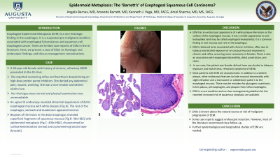
Case Description/Methods: A 58-year-old female with history of chronic, refractory GERD presented to the GI clinic. She reported worsening reflux and heartburn despite being on high-dose proton pump inhibitors. She denied any abdominal pain, nausea, vomiting. She was a non-smoker and denied alcohol use. Her vital signs were normal and physical examination was unremarkable. An upper GI endoscopy revealed abnormal appearance of distal esophageal mucosa with white plaques (Fig A). The rest of the esophagus, stomach and duodenum appeared normal. Biopsies of the lesion in the distal esophagus revealed superficial fragments of squamous mucosa (Fig B- 40x H&E) with epidermoid metaplasia (Fig C- 400x H&E), characterized by surface keratinization (arrow) and a prominent granular layer (bracket).
Discussion: EEM has an endoscopic appearance of a white plaque-like lesion on the surface of the esophageal mucosa. It has a similar appearance to oral leukoplakia (and may be called esophageal leukoplakia); it is a common finding in oral mucosa, but rare in the esophagus. EEM is believed to be associated with chronic irritation, often due to tobacco and alcohol exposure or an unusual mucosal response to chronic acid reflux, occurring more commonly in females. There is also an association with esophageal dysmotility, distal constriction, and stasis. In our case, the patient was female with chronic, refractory symptoms of GERD. Most patients with EEM are asymptomatic. In addition to a whitish plaque, other endoscopic features include mucosal abnormality with slight elevation and a translucent or cobblestone pattern to the esophageal mucosa. EEM is a rare condition and no clear management guidelines for the reported increased risk of squamous neoplasia are available. Little is known about its natural course or risk of malignant progression. Some case reports suggest endoscopic resection. However, most of the literature recommend close follow-up. Further epidemiological and longitudinal studies of EEM are needed.

Disclosures:
Angela Barnes, MD, Amanda Barrett, MD, Kenneth J. Vega, MD, FACG, Amol Sharma, MD, MS, FACG. P3343 - Epidermoid Metaplasia: The 'Barrett's' of Esophageal Squamous Cell Carcinoma?, ACG 2023 Annual Scientific Meeting Abstracts. Vancouver, BC, Canada: American College of Gastroenterology.
Medical College of Georgia at Augusta University, Augusta, GA
Introduction: Esophageal Epidermoid Metaplasia (EEM) is a rare histologic finding in the esophagus. It is a suspected pre-malignant condition associated with esophageal lichen planus and squamous cell esophageal cancer. There are limited case reports of EEM in the GI literature. Here, we present a case of EEM, its histologic and endoscopic findings, and discuss management considerations.
Case Description/Methods: A 58-year-old female with history of chronic, refractory GERD presented to the GI clinic. She reported worsening reflux and heartburn despite being on high-dose proton pump inhibitors. She denied any abdominal pain, nausea, vomiting. She was a non-smoker and denied alcohol use. Her vital signs were normal and physical examination was unremarkable. An upper GI endoscopy revealed abnormal appearance of distal esophageal mucosa with white plaques (Fig A). The rest of the esophagus, stomach and duodenum appeared normal. Biopsies of the lesion in the distal esophagus revealed superficial fragments of squamous mucosa (Fig B- 40x H&E) with epidermoid metaplasia (Fig C- 400x H&E), characterized by surface keratinization (arrow) and a prominent granular layer (bracket).
Discussion: EEM has an endoscopic appearance of a white plaque-like lesion on the surface of the esophageal mucosa. It has a similar appearance to oral leukoplakia (and may be called esophageal leukoplakia); it is a common finding in oral mucosa, but rare in the esophagus. EEM is believed to be associated with chronic irritation, often due to tobacco and alcohol exposure or an unusual mucosal response to chronic acid reflux, occurring more commonly in females. There is also an association with esophageal dysmotility, distal constriction, and stasis. In our case, the patient was female with chronic, refractory symptoms of GERD. Most patients with EEM are asymptomatic. In addition to a whitish plaque, other endoscopic features include mucosal abnormality with slight elevation and a translucent or cobblestone pattern to the esophageal mucosa. EEM is a rare condition and no clear management guidelines for the reported increased risk of squamous neoplasia are available. Little is known about its natural course or risk of malignant progression. Some case reports suggest endoscopic resection. However, most of the literature recommend close follow-up. Further epidemiological and longitudinal studies of EEM are needed.

Figure: Distal esophageal mucosa with white plaques (Fig A).
Superficial fragments of squamous mucosa (Fig B- 40x H&E) with epidermoid metaplasia (Fig C- 400x H&E), characterized by surface keratinization (arrow) and a prominent granular layer (bracket).
Superficial fragments of squamous mucosa (Fig B- 40x H&E) with epidermoid metaplasia (Fig C- 400x H&E), characterized by surface keratinization (arrow) and a prominent granular layer (bracket).
Disclosures:
Angela Barnes indicated no relevant financial relationships.
Amanda Barrett indicated no relevant financial relationships.
Kenneth Vega indicated no relevant financial relationships.
Amol Sharma indicated no relevant financial relationships.
Angela Barnes, MD, Amanda Barrett, MD, Kenneth J. Vega, MD, FACG, Amol Sharma, MD, MS, FACG. P3343 - Epidermoid Metaplasia: The 'Barrett's' of Esophageal Squamous Cell Carcinoma?, ACG 2023 Annual Scientific Meeting Abstracts. Vancouver, BC, Canada: American College of Gastroenterology.
