Tuesday Poster Session
Category: Esophagus
P3344 - An Unusual Cause of Chest Pain: Epiphrenic Diverticulitis
Tuesday, October 24, 2023
10:30 AM - 4:00 PM PT
Location: Exhibit Hall

Has Audio

Megan White, MS, MD
BJC Healthcare
St Louis, MO
Presenting Author(s)
Megan White, MS, MD1, Surachai Amornsawadwattana, MD2, C. Prakash Gyawali, MD, MRCP3
1BJC Healthcare, St. Louis, MO; 2Washington University, St. Louis, MO; 3Washington University School of Medicine, St. Louis, MO
Introduction: An epiphrenic diverticulum (ED) is a pseudodiverticula located within 10 cm of gastroesophageal junction. ED is uncommon and comprises of only 2-4% of all esophageal diverticula. Esophageal motility disorders are present in the majority of patients with ED. The presenting symptoms include heartburn, chest pain, and dysphagia. Treatment is often surgery. Epiphrenic diverticulitis is extremely rare and only a few case reports exist in the literature.
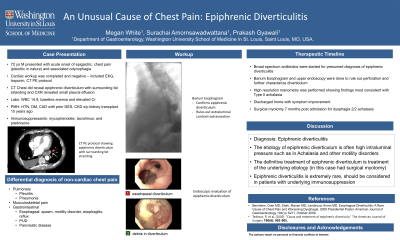
Case Description/Methods: A 72-year-old male with history of hypertension, type 2 diabetes, coronary artery disease, chronic kidney disease status post kidney transplant 15 years ago on chronic immunosuppression presented with 1-day history of chest pain and odynophagia. He denied symptoms of chest pain and dysphagia prior to this admission. Labs were notable for leukocytosis (14,900 mcL) as well as elevated BUN/Cr level. EKG showed sinus bradycardia. Cardiac enzymes were normal. CT PE protocol showed ED with surrounding fat stranding consistent with diverticulitis (figure A). He was started on broad spectrum antibiotics. Inpatient GI service was consulted. A barium esophagram (figure B) was performed and revealed ED without extravasation of contrast. He underwent an upper endoscopy that demonstrated a diverticulum with large opening 38 cm from the incisors (figure C) with food and pus. Then a high resolution esophageal manometry was performed and revealed panesophageal pressure compartmentalization, most similar to a type 2 achalasia. He was evaluated by thoracic surgery during admission as well. He was started on broad spectrum antibiotics and discharged home with improved pain. He underwent surgical myotomy 7 months later when he began to have dysphagia symptoms. His operation went well and on follow up he reported resolution of pain and dysphagia symptoms.
Discussion: Here we report an unusual complication of ED, epiphrenic diverticulitis. There are only a few cases of epiphrenic diverticulitis in the literature and in most cases were treated with antibiotics. ED is usually associated with esophageal motility disorder or increased pressure at the gastroesophageal junction such as from external compression. Therefore, once a diagnosis of ED is confirmed, a workup of underlying etiology should be pursued. This is typically done with an upper endoscopy followed by a high resolution manometry if negative. Treatment of ED is aimed at the underlying disorder and should be performed in symptomatic patients or in patients with large diverticula.

Disclosures:
Megan White, MS, MD1, Surachai Amornsawadwattana, MD2, C. Prakash Gyawali, MD, MRCP3. P3344 - An Unusual Cause of Chest Pain: Epiphrenic Diverticulitis, ACG 2023 Annual Scientific Meeting Abstracts. Vancouver, BC, Canada: American College of Gastroenterology.
1BJC Healthcare, St. Louis, MO; 2Washington University, St. Louis, MO; 3Washington University School of Medicine, St. Louis, MO
Introduction: An epiphrenic diverticulum (ED) is a pseudodiverticula located within 10 cm of gastroesophageal junction. ED is uncommon and comprises of only 2-4% of all esophageal diverticula. Esophageal motility disorders are present in the majority of patients with ED. The presenting symptoms include heartburn, chest pain, and dysphagia. Treatment is often surgery. Epiphrenic diverticulitis is extremely rare and only a few case reports exist in the literature.
Case Description/Methods: A 72-year-old male with history of hypertension, type 2 diabetes, coronary artery disease, chronic kidney disease status post kidney transplant 15 years ago on chronic immunosuppression presented with 1-day history of chest pain and odynophagia. He denied symptoms of chest pain and dysphagia prior to this admission. Labs were notable for leukocytosis (14,900 mcL) as well as elevated BUN/Cr level. EKG showed sinus bradycardia. Cardiac enzymes were normal. CT PE protocol showed ED with surrounding fat stranding consistent with diverticulitis (figure A). He was started on broad spectrum antibiotics. Inpatient GI service was consulted. A barium esophagram (figure B) was performed and revealed ED without extravasation of contrast. He underwent an upper endoscopy that demonstrated a diverticulum with large opening 38 cm from the incisors (figure C) with food and pus. Then a high resolution esophageal manometry was performed and revealed panesophageal pressure compartmentalization, most similar to a type 2 achalasia. He was evaluated by thoracic surgery during admission as well. He was started on broad spectrum antibiotics and discharged home with improved pain. He underwent surgical myotomy 7 months later when he began to have dysphagia symptoms. His operation went well and on follow up he reported resolution of pain and dysphagia symptoms.
Discussion: Here we report an unusual complication of ED, epiphrenic diverticulitis. There are only a few cases of epiphrenic diverticulitis in the literature and in most cases were treated with antibiotics. ED is usually associated with esophageal motility disorder or increased pressure at the gastroesophageal junction such as from external compression. Therefore, once a diagnosis of ED is confirmed, a workup of underlying etiology should be pursued. This is typically done with an upper endoscopy followed by a high resolution manometry if negative. Treatment of ED is aimed at the underlying disorder and should be performed in symptomatic patients or in patients with large diverticula.

Figure: A) CT Chest PE protocol showing epiphrenic diverticulum with surrounding fat stranding, B) barium esophagram showing epiphrenic diverticulum without leak, C) upper endoscopy showing large epiphrenic diverticulum with debris and pus in the lumen
Disclosures:
Megan White indicated no relevant financial relationships.
Surachai Amornsawadwattana indicated no relevant financial relationships.
C. Prakash Gyawali: Diversatek – Consultant, Grant/Research Support. Medtronic – Consultant.
Megan White, MS, MD1, Surachai Amornsawadwattana, MD2, C. Prakash Gyawali, MD, MRCP3. P3344 - An Unusual Cause of Chest Pain: Epiphrenic Diverticulitis, ACG 2023 Annual Scientific Meeting Abstracts. Vancouver, BC, Canada: American College of Gastroenterology.
