Tuesday Poster Session
Category: Esophagus
P3353 - Rare Incidence of Granular Cell Tumor in the Esophagus Causing Obstructive and Reflux Symptoms
Tuesday, October 24, 2023
10:30 AM - 4:00 PM PT
Location: Exhibit Hall

Has Audio

Ramnik Gill, MD, MS
New York Presbyterian Brooklyn Methodist Hospital
Brooklyn, NY
Presenting Author(s)
Ramnik K. Gill, MD, MS1, Ranbir Singh, MD2, Irvin Grosman, MD1
1New York Presbyterian Brooklyn Methodist, Brooklyn, NY; 2NYP Brooklyn Methodist, Brookyln, NY
Introduction: We present the case of a patient diagnosed with Granular Cell Tumor in the Esophagus.
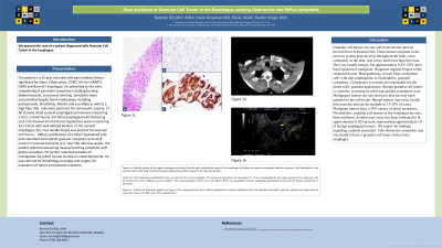
Case Description/Methods: The patient is a 55 year old male with past medical history significant for latent Tuberculosis, COPD, HIV (on HAART), GERD and Barrett’s Esophagus. He presented to the clinic complaining of persistent symptoms including burning abdominal pain, occasional vomiting. Symptoms were uncontrolled despite home medications including pantoprazole, famotidine, Maalox and sucralfate as well as a high fiber diet. Labs were pertinent for normocytic anemia. CT AP showed distal cervical esophageal prominence measuring 1.6cm, a hiatal hernia and distal esophageal wall thickening [2]. EUS showed an intramural, hypoechoic lesion measuring 24 x 13mm with well defined borders in the cervical esophagus [3]. Fine needle biopsy was positive for Granular Cell Tumor - diffuse proliferation of uniform epithelioid cells with abundant eosinophilic granular cytoplasm and small nuclei in muscularis propria [1]. Over the next few weeks, the patient reported worsening nausea/vomiting symptoms with globus sensation. He further reported episodes of constipation for which he was started on metoclopramide. He was referred to hematology-oncology and surgery for evaluation of tumor and possible resection.
Discussion: Granular cell tumors are rare soft tissue tumors that are derived from Schwann Cells. These tumors originate in the nervous system and can arise throughout the body, most commonly in the skin, oral cavity and lower digestive tract. They are usually benign, but approximately 0.5%-2.0% have been reported as malignant. Diagnosis requires biopsy of the suspected lesion. Histopathology reveals large, polygonal cells with pale amphophilic to eosinophilic, granular cytoplasm. Cytoplasmic lysosome are responsible for the tumor cells’ granular appearance. Benign granular cell tumor is common, occurring in soft tissue and the respiratory tract. Malignancy tumors are rare and have thus far only been reported in the soft tissue. Benign tumors may recur locally post excision and can be multiple in 15-20% of cases. Malignant tumors have a 50% chance of distal metastasis. Nevertheless, granular cell tumors of the esophagus are rare; their incidence in endoscopy series has been estimated to be approximately 0.033 percent, representing approximately 1% of benign esophageal tumors. We report our findings regarding a patient presented with obstructive symptoms and was found to have a granular cell tumor in the lower esophagus.

Disclosures:
Ramnik K. Gill, MD, MS1, Ranbir Singh, MD2, Irvin Grosman, MD1. P3353 - Rare Incidence of Granular Cell Tumor in the Esophagus Causing Obstructive and Reflux Symptoms, ACG 2023 Annual Scientific Meeting Abstracts. Vancouver, BC, Canada: American College of Gastroenterology.
1New York Presbyterian Brooklyn Methodist, Brooklyn, NY; 2NYP Brooklyn Methodist, Brookyln, NY
Introduction: We present the case of a patient diagnosed with Granular Cell Tumor in the Esophagus.
Case Description/Methods: The patient is a 55 year old male with past medical history significant for latent Tuberculosis, COPD, HIV (on HAART), GERD and Barrett’s Esophagus. He presented to the clinic complaining of persistent symptoms including burning abdominal pain, occasional vomiting. Symptoms were uncontrolled despite home medications including pantoprazole, famotidine, Maalox and sucralfate as well as a high fiber diet. Labs were pertinent for normocytic anemia. CT AP showed distal cervical esophageal prominence measuring 1.6cm, a hiatal hernia and distal esophageal wall thickening [2]. EUS showed an intramural, hypoechoic lesion measuring 24 x 13mm with well defined borders in the cervical esophagus [3]. Fine needle biopsy was positive for Granular Cell Tumor - diffuse proliferation of uniform epithelioid cells with abundant eosinophilic granular cytoplasm and small nuclei in muscularis propria [1]. Over the next few weeks, the patient reported worsening nausea/vomiting symptoms with globus sensation. He further reported episodes of constipation for which he was started on metoclopramide. He was referred to hematology-oncology and surgery for evaluation of tumor and possible resection.
Discussion: Granular cell tumors are rare soft tissue tumors that are derived from Schwann Cells. These tumors originate in the nervous system and can arise throughout the body, most commonly in the skin, oral cavity and lower digestive tract. They are usually benign, but approximately 0.5%-2.0% have been reported as malignant. Diagnosis requires biopsy of the suspected lesion. Histopathology reveals large, polygonal cells with pale amphophilic to eosinophilic, granular cytoplasm. Cytoplasmic lysosome are responsible for the tumor cells’ granular appearance. Benign granular cell tumor is common, occurring in soft tissue and the respiratory tract. Malignancy tumors are rare and have thus far only been reported in the soft tissue. Benign tumors may recur locally post excision and can be multiple in 15-20% of cases. Malignant tumors have a 50% chance of distal metastasis. Nevertheless, granular cell tumors of the esophagus are rare; their incidence in endoscopy series has been estimated to be approximately 0.033 percent, representing approximately 1% of benign esophageal tumors. We report our findings regarding a patient presented with obstructive symptoms and was found to have a granular cell tumor in the lower esophagus.

Figure: Figure 1a: Mass-like density of the upper esophagus protruding from the right anterolateral
aspect of the esophagus just below the superior esophageal sphincter as seen on CT chest (red
arrow), causing a mild indentation in the posterior wall of the upper trachea and barely
abutting the posterior aspect of the right thyroid lobe.
Figure 1b: Oval intramural (subepithelial) lesion was found in the cervical esophagus (yellow
arrow) during EUS. The lesion was hypoechoic and measured 24 x 13 mm. Sonographically, the
origin appeared to be contiguous with the fourth layer of the esophagus but was exophytic. The
endosonographic borders were well-defined. There was sonographic evidence suggesting
parenchymal invasion into the trachea (manifested by abutment).
Figure 1c: Endoscopic ultrasound guided core biopsy of the esophageal lesion using H&E Stain
shows diffuse proliferation of uniform epithelioid cells with abundant eosinophilic granular
cytoplasm and small nuclei in muscularis propria (blue arrow), consistent with a granular cell
tumor.
Disclosures:
Ramnik Gill indicated no relevant financial relationships.
Ranbir Singh indicated no relevant financial relationships.
Irvin Grosman indicated no relevant financial relationships.
Ramnik K. Gill, MD, MS1, Ranbir Singh, MD2, Irvin Grosman, MD1. P3353 - Rare Incidence of Granular Cell Tumor in the Esophagus Causing Obstructive and Reflux Symptoms, ACG 2023 Annual Scientific Meeting Abstracts. Vancouver, BC, Canada: American College of Gastroenterology.
