Tuesday Poster Session
Category: General Endoscopy
P3425 - A Truly Amorphous Finding: A Case of Amyloid of the Duodenum, a Disease Not to Be Missed
Tuesday, October 24, 2023
10:30 AM - 4:00 PM PT
Location: Exhibit Hall

Has Audio
- MN
Marc Novak, BA
Northwell Health Endoscopy Center of Long Island
Garden City, NY
Presenting Author(s)
Marc Novak, BA, Zachary Marwil, MD
Northwell Health Endoscopy Center of Long Island, Garden City, NY
Introduction: Amyloid of the gastrointestinal tract has a highly variable presentation at diagnosis. Reports state that upwards of 76% of patients with systemic amyloidosis have gastrointestinal involvement at diagnosis. However, 20% of patients will present with only gastrointestinal involvement and no evidence of systemic disease. The gastrointestinal presentation is variable hepatomegaly without splenomegaly , gastrointestinal bleeding in 25-40%, protein losing enteropathy and malabsorption, and dysmotility (gastroparesis or constipation/obstructive symptoms). At diagnosis of GI amyloidosis mucosal infiltration of the small bowel occurs in 100% of patients, stomach and colorectal involvement in >90%, and esophageal involvement in > 70% of patients.
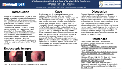
Case Description/Methods: This is a case of a 54 yo woman who presented for evaluation of long-standing reflux and a positive cologuard test. A colonoscopy revealed 3 large polyps ( >1 cm sessile) and 3 diminutive polyps. The large polyps were serrated adenomas, the others were hyperplastic lesions. None contained amyloid. Upper endoscopy revealed LA Grade B esophagitis, a 15 mm submucosal “polyp” at the 2nd portion of the duodenal bulb, and a circumferential soft thickened fold at the apex of the bulb. (figures 1A and 1B). Pathology of the “polyp” and the apical fold revealed submucosal amorphous material that was congo red stain positive, consistent with amyloid. A subsequent bone marrow biopsy did not reveal any plasma cells. Cardiac MRI was also negative; however, SPEP revealed a mild elevation in free kappa and lambda light chains. As the disease appears mild, she does not require therapy at this time and will continue to follow up with GI and hematology for close monitoring.
Discussion: This case highlights the importance of a thorough evaluation of all endoscopic findings, even if incidental in nature. In cases where standard biopsies are non-diagnostic, endoscopic ultrasound with deeper biopsies or other diagnostic modalities might be indicated. In this case, the lesions in the duodenum revealed submucosal deposition of amyloid. While these findings may not explain the patient's presenting symptoms, this case reinforces the need to thoroughly investigate any endoscopic incidental finding. Early diagnosis of amyloid disease will hopefully prevent systemic involvement and improve the overall prognosis.

Disclosures:
Marc Novak, BA, Zachary Marwil, MD. P3425 - A Truly Amorphous Finding: A Case of Amyloid of the Duodenum, a Disease Not to Be Missed, ACG 2023 Annual Scientific Meeting Abstracts. Vancouver, BC, Canada: American College of Gastroenterology.
Northwell Health Endoscopy Center of Long Island, Garden City, NY
Introduction: Amyloid of the gastrointestinal tract has a highly variable presentation at diagnosis. Reports state that upwards of 76% of patients with systemic amyloidosis have gastrointestinal involvement at diagnosis. However, 20% of patients will present with only gastrointestinal involvement and no evidence of systemic disease. The gastrointestinal presentation is variable hepatomegaly without splenomegaly , gastrointestinal bleeding in 25-40%, protein losing enteropathy and malabsorption, and dysmotility (gastroparesis or constipation/obstructive symptoms). At diagnosis of GI amyloidosis mucosal infiltration of the small bowel occurs in 100% of patients, stomach and colorectal involvement in >90%, and esophageal involvement in > 70% of patients.
Case Description/Methods: This is a case of a 54 yo woman who presented for evaluation of long-standing reflux and a positive cologuard test. A colonoscopy revealed 3 large polyps ( >1 cm sessile) and 3 diminutive polyps. The large polyps were serrated adenomas, the others were hyperplastic lesions. None contained amyloid. Upper endoscopy revealed LA Grade B esophagitis, a 15 mm submucosal “polyp” at the 2nd portion of the duodenal bulb, and a circumferential soft thickened fold at the apex of the bulb. (figures 1A and 1B). Pathology of the “polyp” and the apical fold revealed submucosal amorphous material that was congo red stain positive, consistent with amyloid. A subsequent bone marrow biopsy did not reveal any plasma cells. Cardiac MRI was also negative; however, SPEP revealed a mild elevation in free kappa and lambda light chains. As the disease appears mild, she does not require therapy at this time and will continue to follow up with GI and hematology for close monitoring.
Discussion: This case highlights the importance of a thorough evaluation of all endoscopic findings, even if incidental in nature. In cases where standard biopsies are non-diagnostic, endoscopic ultrasound with deeper biopsies or other diagnostic modalities might be indicated. In this case, the lesions in the duodenum revealed submucosal deposition of amyloid. While these findings may not explain the patient's presenting symptoms, this case reinforces the need to thoroughly investigate any endoscopic incidental finding. Early diagnosis of amyloid disease will hopefully prevent systemic involvement and improve the overall prognosis.

Figure:
Figure 1. A: The 15mm submucosal polyp B: Thickened fold of the duodenal bulb
Figure 1. A: The 15mm submucosal polyp B: Thickened fold of the duodenal bulb
Disclosures:
Marc Novak indicated no relevant financial relationships.
Zachary Marwil indicated no relevant financial relationships.
Marc Novak, BA, Zachary Marwil, MD. P3425 - A Truly Amorphous Finding: A Case of Amyloid of the Duodenum, a Disease Not to Be Missed, ACG 2023 Annual Scientific Meeting Abstracts. Vancouver, BC, Canada: American College of Gastroenterology.
