Tuesday Poster Session
Category: General Endoscopy
P3442 - Systemic Mastocytosis
Tuesday, October 24, 2023
10:30 AM - 4:00 PM PT
Location: Exhibit Hall

Has Audio
- KJ
Kendra Jobe, MD
University of Virginia
Charlottesville, VA
Presenting Author(s)
Award: Presidential Poster Award
Kendra Jobe, MD, Rebecca M. Haug, MD, Dennis Kumral, MD
University of Virginia, Charlottesville, VA
Introduction: Mastocytosis is a hematologic disorder characterized by the accumulation of abnormal mast cells in one or more organs. The clinical presentation of systemic mastocytosis varies widely, though 80% present with nonspecific GI manifestations. In adults, systemic disease is likely present if urticaria pigmentosa is noted. Diagnosis is often elusive and can take several years.
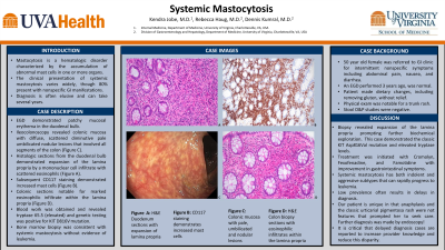
Case Description/Methods: A 50 year old female was referred to GI clinic for intermittent nonspecific GI symptoms including abdominal pain, nausea, and diarrhea. She had no history of anaphylaxis. EGD had been performed for dyspepsia 3 years prior and was normal. At the time the patient made dietary changes including removing dairy products and gluten without relief. Physical exam revealed trunk rash. Repeat EGD, demonstrated patchy mucosal erythema in the duodenal bulb. Ileocolonoscopy revealed colonic mucosa with diffuse, scattered diminutive pale umbilicated nodular lesions that involved all segments of the colon. Histologic sections demonstrated expansion of the lamina propria by a mononuclear cell infiltrate with scattered eosinophils. Colonic sections notable for marked eosinophilic infiltrate within the lamina propria. Blood work was obtained and revealed tryptase 65.5 (elevated) and genetic testing was positive for KIT D816V mutation. Stool O&P studies were negative. Bone marrow biopsy was consistent with systemic mastocytosis without evidence of leukemia. Treatment was initiated with Cromolyn, Fexofenadine, and Famotidine with improvement in gastrointestinal symptoms.
Discussion: Systemic mastocytosis has both indolent and aggressive subtypes that can rapidly progress to leukemia. However the low prevalence often results in delays in diagnosis. Our patient is unique in that anaphylaxis and the classic urticarial pigmentosa rash were not features that prompted her to seek care. Biopsy revealed expansion of the lamina propria prompting further biochemical exploration. This case demonstrated the classic KIT Asp816Val mutation, elevated tryptase levels and responded to antihistamine treatment. Fortunately, she had no evidence of leukemia on bone marrow biopsy. It is critical that delayed diagnosis cases are reported to increase provider knowledge and reduce this disparity.

Disclosures:
Kendra Jobe, MD, Rebecca M. Haug, MD, Dennis Kumral, MD. P3442 - Systemic Mastocytosis, ACG 2023 Annual Scientific Meeting Abstracts. Vancouver, BC, Canada: American College of Gastroenterology.
Kendra Jobe, MD, Rebecca M. Haug, MD, Dennis Kumral, MD
University of Virginia, Charlottesville, VA
Introduction: Mastocytosis is a hematologic disorder characterized by the accumulation of abnormal mast cells in one or more organs. The clinical presentation of systemic mastocytosis varies widely, though 80% present with nonspecific GI manifestations. In adults, systemic disease is likely present if urticaria pigmentosa is noted. Diagnosis is often elusive and can take several years.
Case Description/Methods: A 50 year old female was referred to GI clinic for intermittent nonspecific GI symptoms including abdominal pain, nausea, and diarrhea. She had no history of anaphylaxis. EGD had been performed for dyspepsia 3 years prior and was normal. At the time the patient made dietary changes including removing dairy products and gluten without relief. Physical exam revealed trunk rash. Repeat EGD, demonstrated patchy mucosal erythema in the duodenal bulb. Ileocolonoscopy revealed colonic mucosa with diffuse, scattered diminutive pale umbilicated nodular lesions that involved all segments of the colon. Histologic sections demonstrated expansion of the lamina propria by a mononuclear cell infiltrate with scattered eosinophils. Colonic sections notable for marked eosinophilic infiltrate within the lamina propria. Blood work was obtained and revealed tryptase 65.5 (elevated) and genetic testing was positive for KIT D816V mutation. Stool O&P studies were negative. Bone marrow biopsy was consistent with systemic mastocytosis without evidence of leukemia. Treatment was initiated with Cromolyn, Fexofenadine, and Famotidine with improvement in gastrointestinal symptoms.
Discussion: Systemic mastocytosis has both indolent and aggressive subtypes that can rapidly progress to leukemia. However the low prevalence often results in delays in diagnosis. Our patient is unique in that anaphylaxis and the classic urticarial pigmentosa rash were not features that prompted her to seek care. Biopsy revealed expansion of the lamina propria prompting further biochemical exploration. This case demonstrated the classic KIT Asp816Val mutation, elevated tryptase levels and responded to antihistamine treatment. Fortunately, she had no evidence of leukemia on bone marrow biopsy. It is critical that delayed diagnosis cases are reported to increase provider knowledge and reduce this disparity.

Figure: A. H&E Duodenum sections with expansion of lamina propria.
B. CD117 staining demonstrates increased mast cells.
C. Colonic mucosa with pale, umbilicated and nodular lesions.
D. H&E Colon biopsy sections with eosinophilic infiltrates within the lamina propria.
B. CD117 staining demonstrates increased mast cells.
C. Colonic mucosa with pale, umbilicated and nodular lesions.
D. H&E Colon biopsy sections with eosinophilic infiltrates within the lamina propria.
Disclosures:
Kendra Jobe indicated no relevant financial relationships.
Rebecca Haug indicated no relevant financial relationships.
Dennis Kumral indicated no relevant financial relationships.
Kendra Jobe, MD, Rebecca M. Haug, MD, Dennis Kumral, MD. P3442 - Systemic Mastocytosis, ACG 2023 Annual Scientific Meeting Abstracts. Vancouver, BC, Canada: American College of Gastroenterology.


