Tuesday Poster Session
Category: General Endoscopy
P3443 - Video Capsule Endoscopy: Unlikely Retention in an Inguinal Hernia With a Novel Method for Noninvasive Removal
Tuesday, October 24, 2023
10:30 AM - 4:00 PM PT
Location: Exhibit Hall

Has Audio

Hannah W. Fiske, MD
Brown University/Rhode Island Hospital
Providence, RI
Presenting Author(s)
Hannah W. Fiske, MD1, Averill Guo, MD2, Harlan Rich, MD3
1Brown University/Rhode Island Hospital, Providence, RI; 2Brown University/Warren Alpert Medical School, Providence, RI; 3Warren Alpert Medical School of Brown University, Providence, RI
Introduction: Video capsule endoscopy (VCE) utilizes a pill-sized wireless camera to record images throughout the GI tract. Since its FDA approval in 2001, VCE has gained popularity due to its noninvasive nature and superior sensitivity for detecting abnormalities in the small intestine. Though complications are rare, if present they are often related to capsule retention (1-3% of cases), most commonly caused by small bowel obstruction (SBO) or Crohn’s Disease. Here we present a case of capsule retention in an inguinal hernia. This is only the second known reported case of retention in a hernia, and the first in an inguinal hernia.
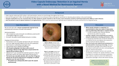
Case Description/Methods: A 58-year-old male with a known asymptomatic inguinal hernia presented with shortness of breath and weakness. GI was consulted for acute microcytic iron deficiency anemia with a hemoglobin (Hgb) of 6.1g/dl. The patient denied abdominal pain or melena. He received two units of packed red blood cells and two units of IV iron; Hgb remained stable for the rest of his admission. EGD showed a nonbleeding esophageal ulcer, and colonoscopy had no significant pathology. VCE showed scalloping in the ileum and an area of narrowing or fibrosis in the distal ileum beyond which the capsule did not advance. Abdominal CT scan revealed the retained capsule in a loop of distal ileum within the known right inguinal hernia. The hernia was manually reduced and a one-liter IV bag was placed over the hernia with pressure wrappings to maintain reduction over the next 24 hours. The patient was subsequently able to pass the capsule spontaneously.
Discussion: When capsule retention is suspected, it is recommended to allow two weeks of observation prior to performing endoscopic or surgical removal. 35-50% of patients pass retained capsules spontaneously within two weeks. Leaving a capsule in the small bowel can result in delayed complications including perforation or SBO. In a previously reported case of capsule retention in an umbilical hernia, hernioplasty was required to achieve eventual capsule expulsion. Here, the retained capsule was successfully dislodged from the herniated ileum using pressure wrappings, avoiding the need for invasive intervention. While a known anatomic hernia is not currently considered a risk factor for capsule retention, it may be prudent to consider pre-procedure evaluation with a patency capsule to assess the likelihood of spontaneous passage in those with known hernias.

Disclosures:
Hannah W. Fiske, MD1, Averill Guo, MD2, Harlan Rich, MD3. P3443 - Video Capsule Endoscopy: Unlikely Retention in an Inguinal Hernia With a Novel Method for Noninvasive Removal, ACG 2023 Annual Scientific Meeting Abstracts. Vancouver, BC, Canada: American College of Gastroenterology.
1Brown University/Rhode Island Hospital, Providence, RI; 2Brown University/Warren Alpert Medical School, Providence, RI; 3Warren Alpert Medical School of Brown University, Providence, RI
Introduction: Video capsule endoscopy (VCE) utilizes a pill-sized wireless camera to record images throughout the GI tract. Since its FDA approval in 2001, VCE has gained popularity due to its noninvasive nature and superior sensitivity for detecting abnormalities in the small intestine. Though complications are rare, if present they are often related to capsule retention (1-3% of cases), most commonly caused by small bowel obstruction (SBO) or Crohn’s Disease. Here we present a case of capsule retention in an inguinal hernia. This is only the second known reported case of retention in a hernia, and the first in an inguinal hernia.
Case Description/Methods: A 58-year-old male with a known asymptomatic inguinal hernia presented with shortness of breath and weakness. GI was consulted for acute microcytic iron deficiency anemia with a hemoglobin (Hgb) of 6.1g/dl. The patient denied abdominal pain or melena. He received two units of packed red blood cells and two units of IV iron; Hgb remained stable for the rest of his admission. EGD showed a nonbleeding esophageal ulcer, and colonoscopy had no significant pathology. VCE showed scalloping in the ileum and an area of narrowing or fibrosis in the distal ileum beyond which the capsule did not advance. Abdominal CT scan revealed the retained capsule in a loop of distal ileum within the known right inguinal hernia. The hernia was manually reduced and a one-liter IV bag was placed over the hernia with pressure wrappings to maintain reduction over the next 24 hours. The patient was subsequently able to pass the capsule spontaneously.
Discussion: When capsule retention is suspected, it is recommended to allow two weeks of observation prior to performing endoscopic or surgical removal. 35-50% of patients pass retained capsules spontaneously within two weeks. Leaving a capsule in the small bowel can result in delayed complications including perforation or SBO. In a previously reported case of capsule retention in an umbilical hernia, hernioplasty was required to achieve eventual capsule expulsion. Here, the retained capsule was successfully dislodged from the herniated ileum using pressure wrappings, avoiding the need for invasive intervention. While a known anatomic hernia is not currently considered a risk factor for capsule retention, it may be prudent to consider pre-procedure evaluation with a patency capsule to assess the likelihood of spontaneous passage in those with known hernias.

Figure: Figure 1:
(a-b) Images from the CT, axial (a) and coronal (b) planes, showing a large right inguinal hernia containing both large and small bowel as well as the ileocecal valve; the retained capsule endoscopy (arrow) is seen within a loop of distal ileum within the right inguinal hernia just proximal to the ileocecal valve; no evidence of obstruction or perforation is noted.
(c) Capsule study showing the narrowing at the base of the hernia with surrounding erythema; capsule was unable to bypass the pictured section of the bowel.
Disclosures:
Hannah Fiske indicated no relevant financial relationships.
Averill Guo indicated no relevant financial relationships.
Harlan Rich indicated no relevant financial relationships.
Hannah W. Fiske, MD1, Averill Guo, MD2, Harlan Rich, MD3. P3443 - Video Capsule Endoscopy: Unlikely Retention in an Inguinal Hernia With a Novel Method for Noninvasive Removal, ACG 2023 Annual Scientific Meeting Abstracts. Vancouver, BC, Canada: American College of Gastroenterology.
