Tuesday Poster Session
Category: GI Bleeding
P3471 - Schistosoma Japonicum Unusually Presenting as a Large Ileocecal Mass: A Case Report
Tuesday, October 24, 2023
10:30 AM - 4:00 PM PT
Location: Exhibit Hall

Has Audio
- JC
Jessa Marie Chua, MD, DPCP
University of Santo Tomas Hospital
Tacloban, Leyte, Philippines
Presenting Author(s)
Jessa Marie Chua, MD, DPCP
University of Santo Tomas Hospital, Tacloban, Leyte, Philippines
Introduction: Schistosomiasis is one of the most neglected tropical parasitic infections in the Philippines. Infection occurs when the larval forms, released by freshwater snails penetrate the skin during contact with infested water. The disease process is due to the Schistosoma eggs which may travel to the different organ systems thru splanchnic circulation causing a wide variety of clinical manifestations that may imitate other gastrointestinal diseases.
Case Description/Methods: A 26-year old previously healthy Filipino male presenting with sudden onset hematochezia associated with crampy generalized abdominal pain. The patient has no known co-morbidities and has no previous work-up, prophylaxis nor treatment for schistosomiasis. Physical examination revealed no signs of hemodynamic instability, his abdomen was soft, non-tender, with hyperactive bowel sounds, and no masses were palpated. Digital rectal exam only showed fresh blood on examining finger. Blood and stool laboratory examinations were all normal, while ultrasound of the whole abdomen only revealed fatty liver. Colonoscopy done showed a large ileocecal mass. The patient then was initially managed as gastrointestinal tuberculosis due to the location of the mass and the endemicity of tuberculosis in the Philippines. However, due to non-improvement of symptoms, surgical management was eventually done. Final result of histopathology examination revealed Schistosomiasis japonicum ova deposits in the colonic mucosa suggesting intestinal schistosomiasis infection.
Discussion: Schistosomiasis may result into unusual presentations due to the migration of eggs to various organs. Intestinal involvement may occur, and the pathogenesis involves host immune responses to the migrating eggs leading to chronic ulceration and granuloma formation. This will eventually lead into acute to chronic colitis associated with bowel wall thickening and in rare instances, may result into colonic mass formation. Diagnosis is done by detection of eggs in the stool as well as biopsy samples. Treatment is usually medical, using Praziquantel, and is achieved by eradicating the adult worm therefore reducing the production of Schistosoma eggs. Surgical intervention is usually unnecessary however may be indicated in certain cases such as low rectal polyps that are within reach, intestinal obstruction, failure to control the clinical manifestations despite adequate treatment, lack of morphological features suggestive of intestinal schistosomiasis and a high suspicion for malignancy.

Disclosures:
Jessa Marie Chua, MD, DPCP. P3471 - Schistosoma Japonicum Unusually Presenting as a Large Ileocecal Mass: A Case Report, ACG 2023 Annual Scientific Meeting Abstracts. Vancouver, BC, Canada: American College of Gastroenterology.
University of Santo Tomas Hospital, Tacloban, Leyte, Philippines
Introduction: Schistosomiasis is one of the most neglected tropical parasitic infections in the Philippines. Infection occurs when the larval forms, released by freshwater snails penetrate the skin during contact with infested water. The disease process is due to the Schistosoma eggs which may travel to the different organ systems thru splanchnic circulation causing a wide variety of clinical manifestations that may imitate other gastrointestinal diseases.
Case Description/Methods: A 26-year old previously healthy Filipino male presenting with sudden onset hematochezia associated with crampy generalized abdominal pain. The patient has no known co-morbidities and has no previous work-up, prophylaxis nor treatment for schistosomiasis. Physical examination revealed no signs of hemodynamic instability, his abdomen was soft, non-tender, with hyperactive bowel sounds, and no masses were palpated. Digital rectal exam only showed fresh blood on examining finger. Blood and stool laboratory examinations were all normal, while ultrasound of the whole abdomen only revealed fatty liver. Colonoscopy done showed a large ileocecal mass. The patient then was initially managed as gastrointestinal tuberculosis due to the location of the mass and the endemicity of tuberculosis in the Philippines. However, due to non-improvement of symptoms, surgical management was eventually done. Final result of histopathology examination revealed Schistosomiasis japonicum ova deposits in the colonic mucosa suggesting intestinal schistosomiasis infection.
Discussion: Schistosomiasis may result into unusual presentations due to the migration of eggs to various organs. Intestinal involvement may occur, and the pathogenesis involves host immune responses to the migrating eggs leading to chronic ulceration and granuloma formation. This will eventually lead into acute to chronic colitis associated with bowel wall thickening and in rare instances, may result into colonic mass formation. Diagnosis is done by detection of eggs in the stool as well as biopsy samples. Treatment is usually medical, using Praziquantel, and is achieved by eradicating the adult worm therefore reducing the production of Schistosoma eggs. Surgical intervention is usually unnecessary however may be indicated in certain cases such as low rectal polyps that are within reach, intestinal obstruction, failure to control the clinical manifestations despite adequate treatment, lack of morphological features suggestive of intestinal schistosomiasis and a high suspicion for malignancy.

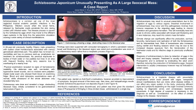
Figure: Colonoscopy showing a large, obstructing mass in the ileocecal area
Disclosures:
Jessa Marie Chua indicated no relevant financial relationships.
Jessa Marie Chua, MD, DPCP. P3471 - Schistosoma Japonicum Unusually Presenting as a Large Ileocecal Mass: A Case Report, ACG 2023 Annual Scientific Meeting Abstracts. Vancouver, BC, Canada: American College of Gastroenterology.

