Tuesday Poster Session
Category: GI Bleeding
P3480 - Spontaneous Omental Bleed Causing Acute Abdomen in Child: A Case Report and Review of Literature
Tuesday, October 24, 2023
10:30 AM - 4:00 PM PT
Location: Exhibit Hall

Has Audio

Chelsie N. Walters, MBBCh
Prince Charles Hospital
Merthyr Tyfdil, Wales, United Kingdom
Presenting Author(s)
Chelsie N. Walters, MBBCh, Ahsan Naqvi, MBBS, MRCS, Shafiq Ahmad Chughtai, MBBS, MRCS, FRCS, MSc
Prince Charles Hospital, Merthyr Tyfdil, Wales, United Kingdom
Introduction: A common condition presenting in children as acute right sided lower abdominal pain is acute appendicitis. Acute appendicitis can be a diagnostic challenge, as presentation can be diverse and misleading.1 Clinical judgement is fundamental in the diagnosis with some support from imaging. However, the poor negative diagnostic value of blood tests and ultrasound scans along with radiation exposure involved in CT scanning make these tests sub optimum for routine use. Often when suspicion is high, a diagnostic laparoscopy is required to confirm the diagnosis. 2
Case Description/Methods: A 12-year-old boy presented with a 1-day history of acute onset vague generalised abdominal pain, shifting to the right lower quadrant. Examination revealed a soft abdomen and localised tenderness in right iliac fossa. His bloods showed normal haemoglobin and white cell count. CRP was mildly elevated, 12mg/L(1-5mg/L) . An ultrasound scan was requested and he was kept in hospital for observation.
Patient was reviewed in ward round after 24 hrs. He was more tender with localised guarding in right iliac fossa. Ultrasound of abdomen was normal. His Padiatric appendicitis score (PAS) was 3. During laparoscopy, appendix was found to be normal, however, blood was found in pelvis and right iliac fossa (150-200 ml). In addition, 4 x 4 cm mass was found within omentum covered with clots. This mass was excised, appendix was removed and peritoneal cavity was washed with saline. The patient went home the following day. Histopathology revealed omental fat with extensive areas of vascular congestion and haemorrhage. Appendix was normal.
Discussion: Right iliac fossa (RIF) pain is one of the most common presentations to the acute surgical take. 4 Common casues mimicking acute appendicitis include, mesenteric adenitis, urinary tract infection or gastroenteritis. 5 Causes of omental haemorrhage includes trauma, aneurysm, malignancy, or vasculitis 6: when the cause is unknown it is classified as idiopathic omental haemorrhage. 7
A search of Ovid Medline data for English language papers revealed only 19 cases of spontaneous omental bleeding.
Only one case of spontaneous omental bleed is reported in pedriatric population.8 All reported cases of omental haemorrhage presented as acute abdominal pain. It appears to be more common in males than in females. This case highlights that spontaneous omental haemorrhage can be a cause of RIF pain 8,9,10,11 and may mimic the symptoms of appendicitis in the pediatric age group.

Disclosures:
Chelsie N. Walters, MBBCh, Ahsan Naqvi, MBBS, MRCS, Shafiq Ahmad Chughtai, MBBS, MRCS, FRCS, MSc. P3480 - Spontaneous Omental Bleed Causing Acute Abdomen in Child: A Case Report and Review of Literature, ACG 2023 Annual Scientific Meeting Abstracts. Vancouver, BC, Canada: American College of Gastroenterology.
Prince Charles Hospital, Merthyr Tyfdil, Wales, United Kingdom
Introduction: A common condition presenting in children as acute right sided lower abdominal pain is acute appendicitis. Acute appendicitis can be a diagnostic challenge, as presentation can be diverse and misleading.1 Clinical judgement is fundamental in the diagnosis with some support from imaging. However, the poor negative diagnostic value of blood tests and ultrasound scans along with radiation exposure involved in CT scanning make these tests sub optimum for routine use. Often when suspicion is high, a diagnostic laparoscopy is required to confirm the diagnosis. 2
Case Description/Methods: A 12-year-old boy presented with a 1-day history of acute onset vague generalised abdominal pain, shifting to the right lower quadrant. Examination revealed a soft abdomen and localised tenderness in right iliac fossa. His bloods showed normal haemoglobin and white cell count. CRP was mildly elevated, 12mg/L(1-5mg/L) . An ultrasound scan was requested and he was kept in hospital for observation.
Patient was reviewed in ward round after 24 hrs. He was more tender with localised guarding in right iliac fossa. Ultrasound of abdomen was normal. His Padiatric appendicitis score (PAS) was 3. During laparoscopy, appendix was found to be normal, however, blood was found in pelvis and right iliac fossa (150-200 ml). In addition, 4 x 4 cm mass was found within omentum covered with clots. This mass was excised, appendix was removed and peritoneal cavity was washed with saline. The patient went home the following day. Histopathology revealed omental fat with extensive areas of vascular congestion and haemorrhage. Appendix was normal.
Discussion: Right iliac fossa (RIF) pain is one of the most common presentations to the acute surgical take. 4 Common casues mimicking acute appendicitis include, mesenteric adenitis, urinary tract infection or gastroenteritis. 5 Causes of omental haemorrhage includes trauma, aneurysm, malignancy, or vasculitis 6: when the cause is unknown it is classified as idiopathic omental haemorrhage. 7
A search of Ovid Medline data for English language papers revealed only 19 cases of spontaneous omental bleeding.
Only one case of spontaneous omental bleed is reported in pedriatric population.8 All reported cases of omental haemorrhage presented as acute abdominal pain. It appears to be more common in males than in females. This case highlights that spontaneous omental haemorrhage can be a cause of RIF pain 8,9,10,11 and may mimic the symptoms of appendicitis in the pediatric age group.

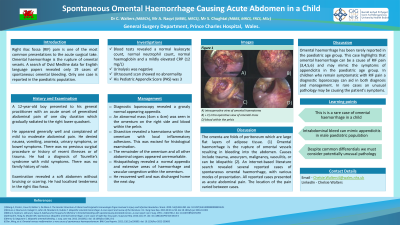
Figure: Diagnostic Laproscopy. Image A,B and C - Intra Op View of Omental Mass. D) Blood in Pelvis
Disclosures:
Chelsie Walters indicated no relevant financial relationships.
Ahsan Naqvi indicated no relevant financial relationships.
Shafiq Ahmad Chughtai indicated no relevant financial relationships.
Chelsie N. Walters, MBBCh, Ahsan Naqvi, MBBS, MRCS, Shafiq Ahmad Chughtai, MBBS, MRCS, FRCS, MSc. P3480 - Spontaneous Omental Bleed Causing Acute Abdomen in Child: A Case Report and Review of Literature, ACG 2023 Annual Scientific Meeting Abstracts. Vancouver, BC, Canada: American College of Gastroenterology.

