Tuesday Poster Session
Category: GI Bleeding
P3492 - A Rare Case of Colonic Varices From Extrinsic Compression of the IVC
Tuesday, October 24, 2023
10:30 AM - 4:00 PM PT
Location: Exhibit Hall

Has Audio
- KG
Kapil Gupta, MD
Rutgers Robert Wood Johnson Medical School
New Brunswick, NJ
Presenting Author(s)
Kapil Gupta, MD, Matthew Pelton, MD, Anish V. Patel, MD
Rutgers Robert Wood Johnson Medical School, New Brunswick, NJ
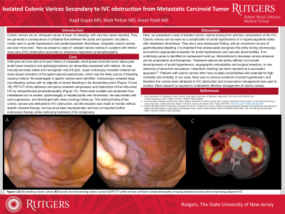
Introduction: Colonic varices are an infrequent cause of lower GI bleeding, with few cases reported. Colonic varices are found in a few conditions, including portal hypertension and portal/mesenteric thrombosis. Only few cases were identified in patients with IVC obstruction. We presented a case of colonic varices arising from extrinsic compression of the IVC.
Case Description/Methods: Patient is a 56-year-old man with PMH of metastatic small bowel carcinoid tumor with a history of surgical gastrojejunostomy with conversion to Roux-en-Y due to prior anastomotic ulcer bleeding who presented for multiple episodes of hematochezia. The patient was initially diagnosed with small bowel NET with small bowel resection and gastrojejunostomy with a primary tumor left in place. He had initially presented to our hospital for melena, which was believed to be from gastrojejunostomy anastomotic ulcers, which required surgical revision to a Roux-en-Y. For 2 years, he had not had further episodes until he began to have intermittent episodes of rectal bleeding, which were intermittent and did not require inpatient admission. For his NET, he had been maintained on medical therapy but, unfortunately, was recently diagnosed with metastatic disease with CT chest, abdomen, and pelvis concerning extrinsic compression of the IVC but patent hepatic/splenic vasculature. He presented to the clinic and was scheduled for endoscopic procedures. The upper endoscopy was notable for congested, erythematous mucosa at the gastrojejunostomy, but no endoscopic finding to explain rectal bleeding. Although with poor preparation, the colonoscopy was notable for congested mucosa at the ileocecal valve, rectal varices, and left-sided colonic varices around 60 cm from the anal verge. After the colonoscopy, the decision was made to monitor him as it was concluded that the compression of the IVC is the primary etiology of the colonic varices. He has remained asymptomatic and has not required further endoscopic therapy while continuing treatment of his NET.
Discussion: Colonic varices are a rare complication of portal venous congestion/portal hypertension. In this case, extrinsic compression of the IVC caused the large varices in the left colon (along with rectal varices). Most patients with colonic varices often have multiple comorbidities and have the potential for high morbidity and mortality. Treatment options must be defined, and more research is required to understand effective control.

Disclosures:
Kapil Gupta, MD, Matthew Pelton, MD, Anish V. Patel, MD. P3492 - A Rare Case of Colonic Varices From Extrinsic Compression of the IVC, ACG 2023 Annual Scientific Meeting Abstracts. Vancouver, BC, Canada: American College of Gastroenterology.
Rutgers Robert Wood Johnson Medical School, New Brunswick, NJ
Introduction: Colonic varices are an infrequent cause of lower GI bleeding, with few cases reported. Colonic varices are found in a few conditions, including portal hypertension and portal/mesenteric thrombosis. Only few cases were identified in patients with IVC obstruction. We presented a case of colonic varices arising from extrinsic compression of the IVC.
Case Description/Methods: Patient is a 56-year-old man with PMH of metastatic small bowel carcinoid tumor with a history of surgical gastrojejunostomy with conversion to Roux-en-Y due to prior anastomotic ulcer bleeding who presented for multiple episodes of hematochezia. The patient was initially diagnosed with small bowel NET with small bowel resection and gastrojejunostomy with a primary tumor left in place. He had initially presented to our hospital for melena, which was believed to be from gastrojejunostomy anastomotic ulcers, which required surgical revision to a Roux-en-Y. For 2 years, he had not had further episodes until he began to have intermittent episodes of rectal bleeding, which were intermittent and did not require inpatient admission. For his NET, he had been maintained on medical therapy but, unfortunately, was recently diagnosed with metastatic disease with CT chest, abdomen, and pelvis concerning extrinsic compression of the IVC but patent hepatic/splenic vasculature. He presented to the clinic and was scheduled for endoscopic procedures. The upper endoscopy was notable for congested, erythematous mucosa at the gastrojejunostomy, but no endoscopic finding to explain rectal bleeding. Although with poor preparation, the colonoscopy was notable for congested mucosa at the ileocecal valve, rectal varices, and left-sided colonic varices around 60 cm from the anal verge. After the colonoscopy, the decision was made to monitor him as it was concluded that the compression of the IVC is the primary etiology of the colonic varices. He has remained asymptomatic and has not required further endoscopic therapy while continuing treatment of his NET.
Discussion: Colonic varices are a rare complication of portal venous congestion/portal hypertension. In this case, extrinsic compression of the IVC caused the large varices in the left colon (along with rectal varices). Most patients with colonic varices often have multiple comorbidities and have the potential for high morbidity and mortality. Treatment options must be defined, and more research is required to understand effective control.

Figure: All four pictures were taken during his colonoscopy. The top two and bottom left picture is of the colonic varices at 60 cm from the anal verge. The bottom right picture is of the rectal varices.
Disclosures:
Kapil Gupta indicated no relevant financial relationships.
Matthew Pelton indicated no relevant financial relationships.
Anish Patel indicated no relevant financial relationships.
Kapil Gupta, MD, Matthew Pelton, MD, Anish V. Patel, MD. P3492 - A Rare Case of Colonic Varices From Extrinsic Compression of the IVC, ACG 2023 Annual Scientific Meeting Abstracts. Vancouver, BC, Canada: American College of Gastroenterology.
