Tuesday Poster Session
Category: GI Bleeding
P3519 - Exulceratio Simplex: A Rare and Old-Described Entity as a Cause of Massive GI Bleeding in the Esophagus
Tuesday, October 24, 2023
10:30 AM - 4:00 PM PT
Location: Exhibit Hall

Has Audio
- RM
Rasheed Musa, MD
East Tennessee State University
Johnson City, TN
Presenting Author(s)
Rasheed Musa, MD1, Koushik Sanku, MD1, Usama Abu-Heija, MBBS1, Ahmad Alnasarat, MD2, Saqr Alsakarneh, MD3, Asaiel Makahleh, MD4, Lana Makahleh, MD1, Larry Schmidt, MD5
1East Tennessee State University, Johnson City, TN; 2Wayne State University/Detroit Medical Center/Sinai Grace Hospital, Detroit, MI; 3University of Missouri-Kansas City, Kansas City, MO; 4East Tennesse State University, Johnson City, TN; 5James H. Quillen VA Medical Center, Mountain Home, TN
Introduction: Dieulafoy's lesion, a dilated aberrant submucosal vessel which erodes the overlying epithelium, is a relatively rare but potentially fatal cause of gastrointestinal (Gl) bleeding. The esophagus is a very rare location for the lesion. Here we present a case of massive upper GI bleeding, secondary to this remarkably rare occurrence, which was amendable to endoscopic intervention.
Case Description/Methods: A 67-year-old male with a medical history significant for peripheral arterial disease status post-femoral-popliteal bypass with recent left common femoral endarterectomy on dual-antiplatelet therapy with aspirin and Plavix, diabetes mellitus type 2, hypertension, hypothyroidism, hyperlipidemia, and ongoing tobacco smoking, who developed acute blood loss anemia and hematemesis.
Labs were obtained and revealed hemoglobin of 5.6.He received a total of 8 units of PRBC transfusion prior to endoscopic evaluation due to ongoing hematemesis.
The patient denied any previous history of gastrointestinal bleeding, denied melena, denied NSAID use, denied heartburn, or reflux-related symptoms, denied nausea and vomiting, denied any known history of chronic liver disease or cirrhosis, he denied alcohol consumption, and he admits smoking a half-pack day of cigarettes, and never had any endoscopic evaluation in the past.
Labs showed CBC with hemoglobin of 5.6, normal platelet count, normal INR, and chemistry was unremarkable except for elevated BUN at 44 and creatinine of 1.4.
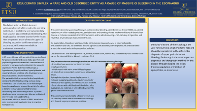
The patient underwent endoscopic evaluation with EGD that showed a visible vessel with active arterial bleeding seen at 30 cm from incisors. Epinephrine injection, hemoclip placement x1 followed by applying gold probe cautery, and lastly hemospray was applied, and hemostasis was achieved.The patient was transferred to a higher level of care at a tertiary center where interventional radiology and thoracic surgery services are available.
The patient eventually did well and no further intervention was required, he was discharged within a week in stable condition.
Discussion: Dieulafoy's lesions of the esophagus are very rare but have a high mortality rate and should be considered in the differential diagnosis of upper gastrointestinal bleeding. Endoscopy is the most important diagnostic and therapeutic method for this disease through clipping the lesion, thermocoagulation or injection of epinephrine, as in our case.

Disclosures:
Rasheed Musa, MD1, Koushik Sanku, MD1, Usama Abu-Heija, MBBS1, Ahmad Alnasarat, MD2, Saqr Alsakarneh, MD3, Asaiel Makahleh, MD4, Lana Makahleh, MD1, Larry Schmidt, MD5. P3519 - Exulceratio Simplex: A Rare and Old-Described Entity as a Cause of Massive GI Bleeding in the Esophagus, ACG 2023 Annual Scientific Meeting Abstracts. Vancouver, BC, Canada: American College of Gastroenterology.
1East Tennessee State University, Johnson City, TN; 2Wayne State University/Detroit Medical Center/Sinai Grace Hospital, Detroit, MI; 3University of Missouri-Kansas City, Kansas City, MO; 4East Tennesse State University, Johnson City, TN; 5James H. Quillen VA Medical Center, Mountain Home, TN
Introduction: Dieulafoy's lesion, a dilated aberrant submucosal vessel which erodes the overlying epithelium, is a relatively rare but potentially fatal cause of gastrointestinal (Gl) bleeding. The esophagus is a very rare location for the lesion. Here we present a case of massive upper GI bleeding, secondary to this remarkably rare occurrence, which was amendable to endoscopic intervention.
Case Description/Methods: A 67-year-old male with a medical history significant for peripheral arterial disease status post-femoral-popliteal bypass with recent left common femoral endarterectomy on dual-antiplatelet therapy with aspirin and Plavix, diabetes mellitus type 2, hypertension, hypothyroidism, hyperlipidemia, and ongoing tobacco smoking, who developed acute blood loss anemia and hematemesis.
Labs were obtained and revealed hemoglobin of 5.6.He received a total of 8 units of PRBC transfusion prior to endoscopic evaluation due to ongoing hematemesis.
The patient denied any previous history of gastrointestinal bleeding, denied melena, denied NSAID use, denied heartburn, or reflux-related symptoms, denied nausea and vomiting, denied any known history of chronic liver disease or cirrhosis, he denied alcohol consumption, and he admits smoking a half-pack day of cigarettes, and never had any endoscopic evaluation in the past.
Labs showed CBC with hemoglobin of 5.6, normal platelet count, normal INR, and chemistry was unremarkable except for elevated BUN at 44 and creatinine of 1.4.
The patient underwent endoscopic evaluation with EGD that showed a visible vessel with active arterial bleeding seen at 30 cm from incisors. Epinephrine injection, hemoclip placement x1 followed by applying gold probe cautery, and lastly hemospray was applied, and hemostasis was achieved.The patient was transferred to a higher level of care at a tertiary center where interventional radiology and thoracic surgery services are available.
The patient eventually did well and no further intervention was required, he was discharged within a week in stable condition.
Discussion: Dieulafoy's lesions of the esophagus are very rare but have a high mortality rate and should be considered in the differential diagnosis of upper gastrointestinal bleeding. Endoscopy is the most important diagnostic and therapeutic method for this disease through clipping the lesion, thermocoagulation or injection of epinephrine, as in our case.

Figure: Dieulafoy's lesions pre and post endoscopic intervention
Disclosures:
Rasheed Musa indicated no relevant financial relationships.
Koushik Sanku indicated no relevant financial relationships.
Usama Abu-Heija indicated no relevant financial relationships.
Ahmad Alnasarat indicated no relevant financial relationships.
Saqr Alsakarneh indicated no relevant financial relationships.
Asaiel Makahleh indicated no relevant financial relationships.
Lana Makahleh indicated no relevant financial relationships.
Larry Schmidt indicated no relevant financial relationships.
Rasheed Musa, MD1, Koushik Sanku, MD1, Usama Abu-Heija, MBBS1, Ahmad Alnasarat, MD2, Saqr Alsakarneh, MD3, Asaiel Makahleh, MD4, Lana Makahleh, MD1, Larry Schmidt, MD5. P3519 - Exulceratio Simplex: A Rare and Old-Described Entity as a Cause of Massive GI Bleeding in the Esophagus, ACG 2023 Annual Scientific Meeting Abstracts. Vancouver, BC, Canada: American College of Gastroenterology.
