Tuesday Poster Session
Category: GI Bleeding
P3525 - Leave No Stone Left Unturned: How Implicit Biases Affect Diagnoses of Dieulafoy’s Lesions
Tuesday, October 24, 2023
10:30 AM - 4:00 PM PT
Location: Exhibit Hall


Anna Lauren G. Winter, MD
University of Mississippi Medical Center
Jackson, MS
Presenting Author(s)
Anna Lauren G. Winter, MD1, Anna Owings, DO1, Ismail Ganim, MD1, David C. Schaefer, MD, PhD2
1University of Mississippi Medical Center, Jackson, MS; 2University of Mississippi Medical Center, Hanover, PA
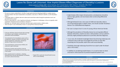
Introduction: The source remains unidentified in 10-20% of upper gastrointestinal bleeding for multiple reasons, including whether the lesion healed before visualization, obscured, or difficult to identify- as with Dieulafoy’s lesions. A Dieulafoy lesion is a dilated, aberrant submucosal vessel that erodes through the epithelium and is not associated with an ulcer. They are responsible for 1-3% of significant GI bleeds, but diagnosis is challenging, so they are arguably under-recognized, making the precise prevalence unknown. Although rare, Dieulafoy lesions have been well-described, predominately in elderly males. Here we report a case of Dieulafoy’s lesion in a young African American female.
Case Description/Methods: A 44-year-old African American female with end-stage renal disease, type 2 diabetes, and hypertension presented to start hemodialysis. She tolerated her first hemodialysis session with no complications. However, during her 2nd session, she became hypotensive, tachycardic, and had two episodes of large-volume hematemesis. She received blood, a proton pump inhibitor, and underwent urgent endoscopy. Endoscopy revealed small ulceration at the GE junction without active bleeding and many blood clots in the fundus. After repositioning for visualization, a bleeding Dieulafoy lesion was spurting blood. An EndoClip was used to achieve hemostasis. Repeat EGD a week later showed healed mucosa with no active bleeding.
Discussion: Implicit biases often impact clinical practice, and physicians should be aware of age, gender, and race biases when constructing differential diagnoses. Although believed to be a rare cause of UGIB and is classically cited as occurring predominately in elderly males, one should consider Dieulafoy lesions in a UGIB differential diagnosis. Our case is unique since it is one of a young African American female. Although the prevalence of Dieulafoy lesions has not typically differed between ethnicities, implicit race bias is still prevalent, and our patient’s ethnicity might predispose physicians to prioritize alternate diagnoses. In our case, the incidental esophageal lesion found during endoscopy was a distractor that could have encouraged the clinician to stop after visualizing the first possible culprit that could cause bleeding. Thankfully, thorough endoscopy found the true culprit under the blood clots in the fundus. In addition to considering implicit biases, it is crucial to leave no stone unturned in clinical practice to provide exceptional patient care.

Disclosures:
Anna Lauren G. Winter, MD1, Anna Owings, DO1, Ismail Ganim, MD1, David C. Schaefer, MD, PhD2. P3525 - Leave No Stone Left Unturned: How Implicit Biases Affect Diagnoses of Dieulafoy’s Lesions, ACG 2023 Annual Scientific Meeting Abstracts. Vancouver, BC, Canada: American College of Gastroenterology.
1University of Mississippi Medical Center, Jackson, MS; 2University of Mississippi Medical Center, Hanover, PA
Introduction: The source remains unidentified in 10-20% of upper gastrointestinal bleeding for multiple reasons, including whether the lesion healed before visualization, obscured, or difficult to identify- as with Dieulafoy’s lesions. A Dieulafoy lesion is a dilated, aberrant submucosal vessel that erodes through the epithelium and is not associated with an ulcer. They are responsible for 1-3% of significant GI bleeds, but diagnosis is challenging, so they are arguably under-recognized, making the precise prevalence unknown. Although rare, Dieulafoy lesions have been well-described, predominately in elderly males. Here we report a case of Dieulafoy’s lesion in a young African American female.
Case Description/Methods: A 44-year-old African American female with end-stage renal disease, type 2 diabetes, and hypertension presented to start hemodialysis. She tolerated her first hemodialysis session with no complications. However, during her 2nd session, she became hypotensive, tachycardic, and had two episodes of large-volume hematemesis. She received blood, a proton pump inhibitor, and underwent urgent endoscopy. Endoscopy revealed small ulceration at the GE junction without active bleeding and many blood clots in the fundus. After repositioning for visualization, a bleeding Dieulafoy lesion was spurting blood. An EndoClip was used to achieve hemostasis. Repeat EGD a week later showed healed mucosa with no active bleeding.
Discussion: Implicit biases often impact clinical practice, and physicians should be aware of age, gender, and race biases when constructing differential diagnoses. Although believed to be a rare cause of UGIB and is classically cited as occurring predominately in elderly males, one should consider Dieulafoy lesions in a UGIB differential diagnosis. Our case is unique since it is one of a young African American female. Although the prevalence of Dieulafoy lesions has not typically differed between ethnicities, implicit race bias is still prevalent, and our patient’s ethnicity might predispose physicians to prioritize alternate diagnoses. In our case, the incidental esophageal lesion found during endoscopy was a distractor that could have encouraged the clinician to stop after visualizing the first possible culprit that could cause bleeding. Thankfully, thorough endoscopy found the true culprit under the blood clots in the fundus. In addition to considering implicit biases, it is crucial to leave no stone unturned in clinical practice to provide exceptional patient care.

Figure: Actively bleeding Dieulafoy's lesion in the fundus.
Disclosures:
Anna Lauren Winter indicated no relevant financial relationships.
Anna Owings indicated no relevant financial relationships.
Ismail Ganim indicated no relevant financial relationships.
David Schaefer indicated no relevant financial relationships.
Anna Lauren G. Winter, MD1, Anna Owings, DO1, Ismail Ganim, MD1, David C. Schaefer, MD, PhD2. P3525 - Leave No Stone Left Unturned: How Implicit Biases Affect Diagnoses of Dieulafoy’s Lesions, ACG 2023 Annual Scientific Meeting Abstracts. Vancouver, BC, Canada: American College of Gastroenterology.
