Monday Poster Session
Category: Biliary/Pancreas
P1474 - What a Fluke! A Case of Intrahepatic Cholangiocarcinoma in a Patient with Schistosoma Infection
Monday, October 23, 2023
10:30 AM - 4:15 PM PT
Location: Exhibit Hall

Has Audio

Arvin N. Tan, MD
University of Hawaii, John A. Burns School of Medicine
Honolulu, HI
Presenting Author(s)
Arvin Tan, MD1, Kevin Edward Nebrejas, MD2, Robert Holm-Kennedy, MD2, Chuong T. Tran, MD3
1University of Hawaii, John A. Burns School of Medicine, Honolulu, HI; 2University of Hawaii, Honolulu, HI; 3Queen's Medical Center, Honolulu, HI
Introduction: Helminthic parasites cause a spectrum of clinical diseases and many carry the potential to develop into malignancies. Schistosoma japonicum has been classified by the IARC as a Group 2B carcinogen: possibly carcinogenic for liver and colorectal carcinoma. To date, only a few cases have been described illustrating the relationship between infection with Schistosoma and cholangiocarcinoma.
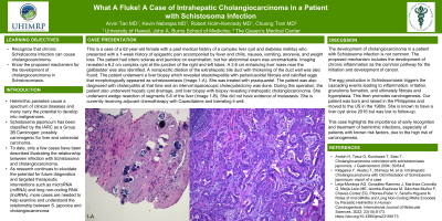
Case Description/Methods: This is a case of a 62-year-old female with a past medical history of a complex liver cyst and diabetes mellitus who presented with a 1-week history of epigastric pain accompanied by fever and chills, nausea, vomiting, anorexia, and weight loss. The patient had icteric sclerae and jaundice on examination, but her abdominal exam was unremarkable. Imaging revealed a 6.2 cm complex cyst at the junction of the right and left lobes. A 2.6 cm enhancing liver mass near the gallbladder was also identified. A nonspecific dilation of the extrahepatic bile duct with thickening of the duct wall was also found. The patient underwent a liver biopsy which revealed steatohepatitis with perisinusoidal fibrosis and calcified eggs that morphologically appeared as schistosomiasis (Image 1-A). She was treated with praziquantel. The patient was also diagnosed with cholecystitis at that time and an interval laparoscopic cholecystectomy was done. During this operation, the patient also underwent hepatic cyst drainage, and liver biopsy with biopsy revealing intrahepatic cholangiocarcinoma. She underwent wedge resection of segments 5-6 of the liver (Image 1-B). She did not have evidence of metastasis. She is currently receiving adjuvant chemotherapy with Capecitabine and tolerating it well.
Discussion: The development of cholangiocarcinoma in a patient with Schistosoma infection is not common. The proposed mechanism includes the development of chronic inflammation as the common pathway for the initiation and development of cancer. The egg production in Schistosomiasis triggers the cascading events leading to inflammation, irritation, granuloma formation, and ultimately fibrosis and hyperplasia. This then promotes carcinogenesis. Our patient was born and raised in the Philippines and moved to the US in the 1980s. She is known to have had a liver cyst since 2016 but was lost to follow-up. This case highlights the importance of early recognition and treatment of helminthic infections, especially of patients with known risk factors, due to the high risk of carcinogenesis.

Disclosures:
Arvin Tan, MD1, Kevin Edward Nebrejas, MD2, Robert Holm-Kennedy, MD2, Chuong T. Tran, MD3. P1474 - What a Fluke! A Case of Intrahepatic Cholangiocarcinoma in a Patient with Schistosoma Infection, ACG 2023 Annual Scientific Meeting Abstracts. Vancouver, BC, Canada: American College of Gastroenterology.
1University of Hawaii, John A. Burns School of Medicine, Honolulu, HI; 2University of Hawaii, Honolulu, HI; 3Queen's Medical Center, Honolulu, HI
Introduction: Helminthic parasites cause a spectrum of clinical diseases and many carry the potential to develop into malignancies. Schistosoma japonicum has been classified by the IARC as a Group 2B carcinogen: possibly carcinogenic for liver and colorectal carcinoma. To date, only a few cases have been described illustrating the relationship between infection with Schistosoma and cholangiocarcinoma.
Case Description/Methods: This is a case of a 62-year-old female with a past medical history of a complex liver cyst and diabetes mellitus who presented with a 1-week history of epigastric pain accompanied by fever and chills, nausea, vomiting, anorexia, and weight loss. The patient had icteric sclerae and jaundice on examination, but her abdominal exam was unremarkable. Imaging revealed a 6.2 cm complex cyst at the junction of the right and left lobes. A 2.6 cm enhancing liver mass near the gallbladder was also identified. A nonspecific dilation of the extrahepatic bile duct with thickening of the duct wall was also found. The patient underwent a liver biopsy which revealed steatohepatitis with perisinusoidal fibrosis and calcified eggs that morphologically appeared as schistosomiasis (Image 1-A). She was treated with praziquantel. The patient was also diagnosed with cholecystitis at that time and an interval laparoscopic cholecystectomy was done. During this operation, the patient also underwent hepatic cyst drainage, and liver biopsy with biopsy revealing intrahepatic cholangiocarcinoma. She underwent wedge resection of segments 5-6 of the liver (Image 1-B). She did not have evidence of metastasis. She is currently receiving adjuvant chemotherapy with Capecitabine and tolerating it well.
Discussion: The development of cholangiocarcinoma in a patient with Schistosoma infection is not common. The proposed mechanism includes the development of chronic inflammation as the common pathway for the initiation and development of cancer. The egg production in Schistosomiasis triggers the cascading events leading to inflammation, irritation, granuloma formation, and ultimately fibrosis and hyperplasia. This then promotes carcinogenesis. Our patient was born and raised in the Philippines and moved to the US in the 1980s. She is known to have had a liver cyst since 2016 but was lost to follow-up. This case highlights the importance of early recognition and treatment of helminthic infections, especially of patients with known risk factors, due to the high risk of carcinogenesis.

Figure: 1-A: Photomicrograph of liver biopsy demonstrating calcified eggs. 1-B: Photomicrograph of liver resection specimen demonstrating cholangiocarcinoma.
Disclosures:
Arvin Tan indicated no relevant financial relationships.
Kevin Edward Nebrejas indicated no relevant financial relationships.
Robert Holm-Kennedy indicated no relevant financial relationships.
Chuong Tran indicated no relevant financial relationships.
Arvin Tan, MD1, Kevin Edward Nebrejas, MD2, Robert Holm-Kennedy, MD2, Chuong T. Tran, MD3. P1474 - What a Fluke! A Case of Intrahepatic Cholangiocarcinoma in a Patient with Schistosoma Infection, ACG 2023 Annual Scientific Meeting Abstracts. Vancouver, BC, Canada: American College of Gastroenterology.
