Monday Poster Session
Category: Biliary/Pancreas
P1486 - A Tale of Two Primaries: Gallbladder Small Cell Carcinoma With Concurrent Adenocarcinoma of the Lung
Monday, October 23, 2023
10:30 AM - 4:15 PM PT
Location: Exhibit Hall

Has Audio
- HI
Humzah Iqbal, MD
University of California San Francisco, Fresno
Fresno, CA
Presenting Author(s)
Humzah Iqbal, MD1, Saranya Sasidharan, MD2, Juliana Yang, MD3
1University of California San Francisco, Fresno, Fresno, CA; 2UCSF Fresno, Fresno, CA; 3University of Texas Medical Branch at Galveston, Galveston, TX
Introduction: Small cell carcinoma (SCC) of the gallbladder is an exceedingly rare clinical entity, accounting for only 0.5% of all gallbladder malignancies. The prognosis is poor, with most patients presenting late in the disease course with the presence of metastases. We present a case of a patient who presented with symptoms concerning for choledocholithiasis, and was found to have SCC of the gallbladder and mucinous adenocarcinoma of the lung.
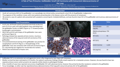
Case Description/Methods: An 81 year old woman presented with fatigue and right upper quadrant pain. She had total bilirubin elevation to 2.5 mg/dL, alkaline phosphatase 734 IU/L, ALT 61 U/L, and AST 68 U/L. Magnetic resonance cholangiopancreatography (MRCP) showed a 6 x 8 cm exophytic mass extending from the gallbladder fundus (Figure a). Computed tomography (CT) imaging revealed pulmonary septal thickening, concerning for possible lymphangitic carcinomatosis. Patient underwent endobronchial ultrasound with fine-needle aspiration (EBUS FNA) and endoscopic ultrasound (EUS) with biopsy of the gallbladder mass (Figure b). Pathological findings were significant for two separate primary tumors, mucinous adenocarcinoma of the lung and small cell carcinoma of the gallbladder. Immunohistochemical staining of the gallbladder was positive for synaptophysin and Ki-67 (Figure c). Histopathology of the gallbladder mass was consistent with small cell carcinoma (Figure d). Pathology of the pulmonary lesions revealed findings consistent with mucinous adenocarcinoma, and negative neuroendocrine immunohistochemical stains. Due to extensive disease burden and poor functional status, patient was transitioned to hospice care.
Discussion: Gallbladder SCC is an extremely rare and aggressive cancer, accounting for 0.5% of all gallbladder cancers. Patients with gallbladder SCC are often diagnosed at late stages with early metastasis to the lymph nodes, liver and lungs. The median survival has been estimated at 4-9 months. Due to the rarity and aggressive nature of gallbladder SCC, standardized treatment regimens are lacking. Our patient’s pulmonary findings initially raised suspicion for a metastatic process. However, she was found to have two primary tumors which proved to be separate entities. Only a handful of cases have reported concurrence of gallbladder SCC with adenocarcinoma, however they primarily describe neoplasms isolated to the gallbladder. To our knowledge, no other case has reported primary adenocarcinoma of the lung with concurrent primary SCC of the gallbladder.

Disclosures:
Humzah Iqbal, MD1, Saranya Sasidharan, MD2, Juliana Yang, MD3. P1486 - A Tale of Two Primaries: Gallbladder Small Cell Carcinoma With Concurrent Adenocarcinoma of the Lung, ACG 2023 Annual Scientific Meeting Abstracts. Vancouver, BC, Canada: American College of Gastroenterology.
1University of California San Francisco, Fresno, Fresno, CA; 2UCSF Fresno, Fresno, CA; 3University of Texas Medical Branch at Galveston, Galveston, TX
Introduction: Small cell carcinoma (SCC) of the gallbladder is an exceedingly rare clinical entity, accounting for only 0.5% of all gallbladder malignancies. The prognosis is poor, with most patients presenting late in the disease course with the presence of metastases. We present a case of a patient who presented with symptoms concerning for choledocholithiasis, and was found to have SCC of the gallbladder and mucinous adenocarcinoma of the lung.
Case Description/Methods: An 81 year old woman presented with fatigue and right upper quadrant pain. She had total bilirubin elevation to 2.5 mg/dL, alkaline phosphatase 734 IU/L, ALT 61 U/L, and AST 68 U/L. Magnetic resonance cholangiopancreatography (MRCP) showed a 6 x 8 cm exophytic mass extending from the gallbladder fundus (Figure a). Computed tomography (CT) imaging revealed pulmonary septal thickening, concerning for possible lymphangitic carcinomatosis. Patient underwent endobronchial ultrasound with fine-needle aspiration (EBUS FNA) and endoscopic ultrasound (EUS) with biopsy of the gallbladder mass (Figure b). Pathological findings were significant for two separate primary tumors, mucinous adenocarcinoma of the lung and small cell carcinoma of the gallbladder. Immunohistochemical staining of the gallbladder was positive for synaptophysin and Ki-67 (Figure c). Histopathology of the gallbladder mass was consistent with small cell carcinoma (Figure d). Pathology of the pulmonary lesions revealed findings consistent with mucinous adenocarcinoma, and negative neuroendocrine immunohistochemical stains. Due to extensive disease burden and poor functional status, patient was transitioned to hospice care.
Discussion: Gallbladder SCC is an extremely rare and aggressive cancer, accounting for 0.5% of all gallbladder cancers. Patients with gallbladder SCC are often diagnosed at late stages with early metastasis to the lymph nodes, liver and lungs. The median survival has been estimated at 4-9 months. Due to the rarity and aggressive nature of gallbladder SCC, standardized treatment regimens are lacking. Our patient’s pulmonary findings initially raised suspicion for a metastatic process. However, she was found to have two primary tumors which proved to be separate entities. Only a handful of cases have reported concurrence of gallbladder SCC with adenocarcinoma, however they primarily describe neoplasms isolated to the gallbladder. To our knowledge, no other case has reported primary adenocarcinoma of the lung with concurrent primary SCC of the gallbladder.

Figure: A: MRCP showing 6 x 8 cm cystic and solid gallbladder mass. B: EUS FNA of gallbladder mass. C: Biopsy of gallbladder mass showing positive neuroendocrine staining. D: Hematoxylin and eosin (H & E) stained slide of gallbladder mass showing findings consistent with small cell carcinoma.
Disclosures:
Humzah Iqbal indicated no relevant financial relationships.
Saranya Sasidharan indicated no relevant financial relationships.
Juliana Yang indicated no relevant financial relationships.
Humzah Iqbal, MD1, Saranya Sasidharan, MD2, Juliana Yang, MD3. P1486 - A Tale of Two Primaries: Gallbladder Small Cell Carcinoma With Concurrent Adenocarcinoma of the Lung, ACG 2023 Annual Scientific Meeting Abstracts. Vancouver, BC, Canada: American College of Gastroenterology.
