Monday Poster Session
Category: Biliary/Pancreas
P1492 - Bouveret Syndrome: A Rare Cause of Duodenal Obstruction
Monday, October 23, 2023
10:30 AM - 4:15 PM PT
Location: Exhibit Hall

Has Audio

Anas Khouri, MD
University of South Alabama
Mobile, AL
Presenting Author(s)
Anas Khouri, MD1, Karan B. Singh, MD1, Kane Edwards, MD2, Samuel B S. Hooks, MD3, Baret T. Bercier, MD2
1University of South Alabama, Mobile, AL; 2USA Health, Mobile, AL; 3University of South Alabama College of Medicine, Mobile, AL
Introduction: Gallstone ileus is a rare complication of cholelithiasis with an incidence of 0.3-0.5%. It occurs when a gallstone causes ileus or obstruction at the ileocecal valve via biliary enteric fistula. Bouveret syndrome accounts for only 1-3% of gallstone ileus, and it occurs when the stone impacts proximally in the duodenum or proximal jejunum causing gastric outlet obstruction. We present a case of Bouveret syndrome in a 70-year-old female who presented with abdominal pain.
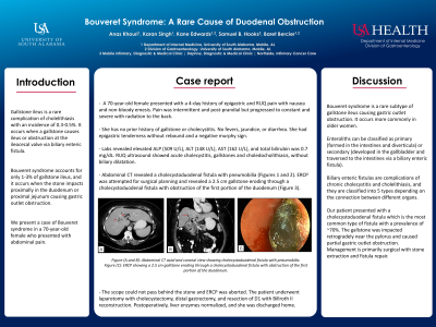
Case Description/Methods: A 70-year-old female presented with a 4-day history of epigastric and RUQ pain with nausea and non-bloody emesis. Pain was intermittent and post-prandial but progressed to constant and severe with radiation to the back. She has no prior history of gallstone or cholecystitis. No fevers, jaundice, or diarrhea. She had epigastric tenderness without rebound and a negative murphy sign. Labs revealed elevated ALP (509 U/L), ALT (148 U/L), AST (162 U/L), and total bilirubin was 0.7 mg/dL. RUQ ultrasound showed acute cholecystitis, gallstones and choledocholithiasis, without biliary dilatation. Abdominal CT revealed a cholecystoduodenal fistula with pneumobilia (Figures 1 and 2). ERCP was attempted for surgical planning and revealed a 2.5 cm gallstone eroding through a cholecystoduodenal fistula with obstruction of the first portion of the duodenum (Figure 3). The scope could not pass behind the stone and ERCP was aborted. The patient underwent laparotomy with cholecystectomy, distal gastrectomy, and resection of D1 with Billroth II reconstruction. Postoperatively, liver enzymes normalized, and she was discharged home.
Discussion: Bouveret syndrome is a rare subtype of gallstone ileus causing gastric outlet obstruction. It occurs more commonly in older women. Enteroliths can be classified as primary (formed in the intestines and diverticula) or secondary (developed in the gallbladder and traversed to the intestines via a biliary enteric fistula). Biliary enteric fistulas are complications of chronic cholecystitis and cholelithiasis, and they are classified into 5 types depending on the connection between different organs. Our patient presented with a cholecystoduodenal fistula which is the most common type of fistula with a prevalence of ~70%. The gallstone was impacted retrogradely near the pylorus and caused partial gastric outlet obstruction. Management is primarily surgical with stone extraction and fistula repair.

Disclosures:
Anas Khouri, MD1, Karan B. Singh, MD1, Kane Edwards, MD2, Samuel B S. Hooks, MD3, Baret T. Bercier, MD2. P1492 - Bouveret Syndrome: A Rare Cause of Duodenal Obstruction, ACG 2023 Annual Scientific Meeting Abstracts. Vancouver, BC, Canada: American College of Gastroenterology.
1University of South Alabama, Mobile, AL; 2USA Health, Mobile, AL; 3University of South Alabama College of Medicine, Mobile, AL
Introduction: Gallstone ileus is a rare complication of cholelithiasis with an incidence of 0.3-0.5%. It occurs when a gallstone causes ileus or obstruction at the ileocecal valve via biliary enteric fistula. Bouveret syndrome accounts for only 1-3% of gallstone ileus, and it occurs when the stone impacts proximally in the duodenum or proximal jejunum causing gastric outlet obstruction. We present a case of Bouveret syndrome in a 70-year-old female who presented with abdominal pain.
Case Description/Methods: A 70-year-old female presented with a 4-day history of epigastric and RUQ pain with nausea and non-bloody emesis. Pain was intermittent and post-prandial but progressed to constant and severe with radiation to the back. She has no prior history of gallstone or cholecystitis. No fevers, jaundice, or diarrhea. She had epigastric tenderness without rebound and a negative murphy sign. Labs revealed elevated ALP (509 U/L), ALT (148 U/L), AST (162 U/L), and total bilirubin was 0.7 mg/dL. RUQ ultrasound showed acute cholecystitis, gallstones and choledocholithiasis, without biliary dilatation. Abdominal CT revealed a cholecystoduodenal fistula with pneumobilia (Figures 1 and 2). ERCP was attempted for surgical planning and revealed a 2.5 cm gallstone eroding through a cholecystoduodenal fistula with obstruction of the first portion of the duodenum (Figure 3). The scope could not pass behind the stone and ERCP was aborted. The patient underwent laparotomy with cholecystectomy, distal gastrectomy, and resection of D1 with Billroth II reconstruction. Postoperatively, liver enzymes normalized, and she was discharged home.
Discussion: Bouveret syndrome is a rare subtype of gallstone ileus causing gastric outlet obstruction. It occurs more commonly in older women. Enteroliths can be classified as primary (formed in the intestines and diverticula) or secondary (developed in the gallbladder and traversed to the intestines via a biliary enteric fistula). Biliary enteric fistulas are complications of chronic cholecystitis and cholelithiasis, and they are classified into 5 types depending on the connection between different organs. Our patient presented with a cholecystoduodenal fistula which is the most common type of fistula with a prevalence of ~70%. The gallstone was impacted retrogradely near the pylorus and caused partial gastric outlet obstruction. Management is primarily surgical with stone extraction and fistula repair.

Figure: Figure (A and B): Abdominal CT axial and coronal view showing cholecystoduodenal fistula with pneumobilia.
Figure (C): ERCP showing a 2.5 cm gallstone eroding through a cholecystoduodenal fistula with obstruction of the first portion of the duodenum.
Figure (C): ERCP showing a 2.5 cm gallstone eroding through a cholecystoduodenal fistula with obstruction of the first portion of the duodenum.
Disclosures:
Anas Khouri indicated no relevant financial relationships.
Karan Singh indicated no relevant financial relationships.
Kane Edwards indicated no relevant financial relationships.
Samuel Hooks indicated no relevant financial relationships.
Baret Bercier indicated no relevant financial relationships.
Anas Khouri, MD1, Karan B. Singh, MD1, Kane Edwards, MD2, Samuel B S. Hooks, MD3, Baret T. Bercier, MD2. P1492 - Bouveret Syndrome: A Rare Cause of Duodenal Obstruction, ACG 2023 Annual Scientific Meeting Abstracts. Vancouver, BC, Canada: American College of Gastroenterology.
