Monday Poster Session
Category: Biliary/Pancreas
P1526 - A Challenging Case of a Rare Entity: Portal Hypertensive Biliopathy in a Patient With Essential Thrombocythemia
Monday, October 23, 2023
10:30 AM - 4:15 PM PT
Location: Exhibit Hall

Has Audio

Hayden Rotramel, MD
Saint Louis University
St. Louis, MO
Presenting Author(s)
Hayden Rotramel, MD, Jay Shah, DO, MSc, Ryan Plunkett, MD, Michelle Baliss, DO, Wissam Kiwan, MD
Saint Louis University, St. Louis, MO
Introduction: Chronic portal vein thrombosis (PVT) is a known cause of noncirrhotic portal hypertension (NCPH) that can result in biliary tract abnormalities and secondary biliary cirrhosis. However, symptomatic portal hypertensive biliopathy (PB) from biliary obstruction is exceedingly rare. Portosystemic shunting is the treatment of choice but may not always be feasible. We present a case of PB in a patient with essential thrombocythemia (ET) in whom portosystemic shunt placement was unsuccessful.
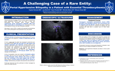
Case Description/Methods: A 66-year-old male with PVT due to ET was admitted with 3 weeks of abdominal pain and jaundice. 3 years prior, he presented with abdominal pain and was found to have extensive right, left, and main PVT with splenic and superior mesenteric vein extension. Elevated platelet counts prompted JAK2-V617F testing, leading to the diagnosis of ET and treatment with hydroxyurea and apixaban. He remained asymptomatic until admission 2 years later with hematemesis. He was found to have esophageal variceal (EV) bleeding due to PVT-induced NCPH managed with band ligation. His course was complicated by cholestatic liver injury (AST 95, ALT 96, ALP 669, bilirubin 4.1). Magnetic resonance cholangiopancreatography (MRCP) showed intrahepatic biliary dilation with severe common bile duct (CBD) narrowing in the porta hepatis near the PVT. ERCP showed a stricture below the hilum with no stones or sludge. Sphincterotomy and plastic stent placement were performed. To further evaluate the indeterminate biliary stricture, endoscopic ultrasound (EUS) was done and revealed dilated collateral vessels around the CBD (likely the stricture etiology), making a diagnosis of PB (Fig 1). TIPS was attempted but was unsuccessful due to extensive vascular fibrosis. Ultimately, he underwent a modified Sugiura procedure for refractory EV bleeding and will be monitored with serial labs to evaluate need for future biliary intervention.
Discussion: PB in the setting of PVT is thought to be caused by vascular and connective tissue neogenesis resulting in angulation, extrinsic bile duct compression, and ischemic strictures. The diagnostic study of choice is MRCP. EUS, while not routinely used for this purpose, can distinguish between CBD varices, stones, and tumors when other modalities are non-diagnostic. Portosystemic shunting to reverse portal biliopathy is indicated in most cases but may not always be feasible as in this case.

Disclosures:
Hayden Rotramel, MD, Jay Shah, DO, MSc, Ryan Plunkett, MD, Michelle Baliss, DO, Wissam Kiwan, MD. P1526 - A Challenging Case of a Rare Entity: Portal Hypertensive Biliopathy in a Patient With Essential Thrombocythemia, ACG 2023 Annual Scientific Meeting Abstracts. Vancouver, BC, Canada: American College of Gastroenterology.
Saint Louis University, St. Louis, MO
Introduction: Chronic portal vein thrombosis (PVT) is a known cause of noncirrhotic portal hypertension (NCPH) that can result in biliary tract abnormalities and secondary biliary cirrhosis. However, symptomatic portal hypertensive biliopathy (PB) from biliary obstruction is exceedingly rare. Portosystemic shunting is the treatment of choice but may not always be feasible. We present a case of PB in a patient with essential thrombocythemia (ET) in whom portosystemic shunt placement was unsuccessful.
Case Description/Methods: A 66-year-old male with PVT due to ET was admitted with 3 weeks of abdominal pain and jaundice. 3 years prior, he presented with abdominal pain and was found to have extensive right, left, and main PVT with splenic and superior mesenteric vein extension. Elevated platelet counts prompted JAK2-V617F testing, leading to the diagnosis of ET and treatment with hydroxyurea and apixaban. He remained asymptomatic until admission 2 years later with hematemesis. He was found to have esophageal variceal (EV) bleeding due to PVT-induced NCPH managed with band ligation. His course was complicated by cholestatic liver injury (AST 95, ALT 96, ALP 669, bilirubin 4.1). Magnetic resonance cholangiopancreatography (MRCP) showed intrahepatic biliary dilation with severe common bile duct (CBD) narrowing in the porta hepatis near the PVT. ERCP showed a stricture below the hilum with no stones or sludge. Sphincterotomy and plastic stent placement were performed. To further evaluate the indeterminate biliary stricture, endoscopic ultrasound (EUS) was done and revealed dilated collateral vessels around the CBD (likely the stricture etiology), making a diagnosis of PB (Fig 1). TIPS was attempted but was unsuccessful due to extensive vascular fibrosis. Ultimately, he underwent a modified Sugiura procedure for refractory EV bleeding and will be monitored with serial labs to evaluate need for future biliary intervention.
Discussion: PB in the setting of PVT is thought to be caused by vascular and connective tissue neogenesis resulting in angulation, extrinsic bile duct compression, and ischemic strictures. The diagnostic study of choice is MRCP. EUS, while not routinely used for this purpose, can distinguish between CBD varices, stones, and tumors when other modalities are non-diagnostic. Portosystemic shunting to reverse portal biliopathy is indicated in most cases but may not always be feasible as in this case.

Figure: Fig 1: Portal Hypertensive Biliopathy on EUS
Disclosures:
Hayden Rotramel indicated no relevant financial relationships.
Jay Shah indicated no relevant financial relationships.
Ryan Plunkett indicated no relevant financial relationships.
Michelle Baliss indicated no relevant financial relationships.
Wissam Kiwan indicated no relevant financial relationships.
Hayden Rotramel, MD, Jay Shah, DO, MSc, Ryan Plunkett, MD, Michelle Baliss, DO, Wissam Kiwan, MD. P1526 - A Challenging Case of a Rare Entity: Portal Hypertensive Biliopathy in a Patient With Essential Thrombocythemia, ACG 2023 Annual Scientific Meeting Abstracts. Vancouver, BC, Canada: American College of Gastroenterology.
