Monday Poster Session
Category: Biliary/Pancreas
P1538 - Role of Endoscopic Ultrasound in a Rare Presentation of a Diffuse Serous Cystadenoma of the Pancreas
Monday, October 23, 2023
10:30 AM - 4:15 PM PT
Location: Exhibit Hall

Has Audio

Ria Kundu, MD
Banner Health
Mesa, AZ
Presenting Author(s)
Aida Rezaie, MD1, Ria Kundu, MD2, Savio Reddymasu, MD3
1Creighton University, Mesa, AZ; 2Banner Health, Mesa, AZ; 3Creighton University, Phoenix, AZ
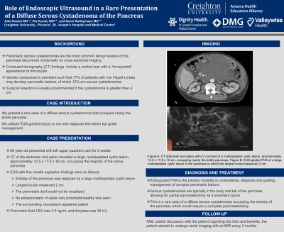
Introduction: Pancreatic serous cystadenomas are the most common benign lesions of the pancreas discovered incidentally on cross-sectional imaging. They typically present in women between the fifth and seventh decade of life in the body or tail of the pancreas. Computed tomography (CT) findings of a serous cystadenoma include a central scar with a “honeycomb” appearance of microcysts. While the etiology is unclear, there may be a genetic component as around 77% of patients with von Hippel-Lindau develop pancreatic lesions, of which 12% are serous cystadenomas. Management depends on patient symptoms and the size of the serous cystadenoma. Surgical resection is usually recommended if it is greater than 4 cm. We present a rare case of a diffuse serous cystadenoma that occupied nearly the entire pancreas, and role of EUS-guided biopsy to not only diagnose this lesion but guide management.
Case Description/Methods: 69 year old presented with left upper quadrant pain for 2 weeks that had since resolved. Computed tomography of the abdomen and pelvis scan showed a large, multiseptated cystic lesion, approximately 12.5 x 17.8 x 16 cm, occupying the majority of the native pancreas. The patient subsequently underwent Endoscopic ultrasound (EUS) with fine needle aspiration which revealed that the entirety of the pancreas was replaced by a large multiseptated cystic lesion in which the largest locule measured 2 cm. The pancreatic duct could not be visualized, no peripancreatic of celiac axis lymphadenopathy was seen and the surrounding vasculature appeared patent. The pancreatic fluid CEA was 3.0 ng/mL and Amylase 35 U/L. The patient was subsequently diagnosed with a diffuse serous cystadenoma of the pancreas.
Discussion: Serous cystadenomas are typically in the body-tail of the pancreas allowing for partial pancreatectomy as a treatment option. EUS-guided FNA is the primary modality to characterize, diagnose and guiding management of complex pancreatic lesions. This is a rare case of a diffuse serous cystadenoma occupying the entirety of the pancreas which would require a complete pancreatectomy. After careful discussion with the patient regarding the risks and benefits, the patient elected to undergo serial imaging with an MRI in 3 months.

Disclosures:
Aida Rezaie, MD1, Ria Kundu, MD2, Savio Reddymasu, MD3. P1538 - Role of Endoscopic Ultrasound in a Rare Presentation of a Diffuse Serous Cystadenoma of the Pancreas, ACG 2023 Annual Scientific Meeting Abstracts. Vancouver, BC, Canada: American College of Gastroenterology.
1Creighton University, Mesa, AZ; 2Banner Health, Mesa, AZ; 3Creighton University, Phoenix, AZ
Introduction: Pancreatic serous cystadenomas are the most common benign lesions of the pancreas discovered incidentally on cross-sectional imaging. They typically present in women between the fifth and seventh decade of life in the body or tail of the pancreas. Computed tomography (CT) findings of a serous cystadenoma include a central scar with a “honeycomb” appearance of microcysts. While the etiology is unclear, there may be a genetic component as around 77% of patients with von Hippel-Lindau develop pancreatic lesions, of which 12% are serous cystadenomas. Management depends on patient symptoms and the size of the serous cystadenoma. Surgical resection is usually recommended if it is greater than 4 cm. We present a rare case of a diffuse serous cystadenoma that occupied nearly the entire pancreas, and role of EUS-guided biopsy to not only diagnose this lesion but guide management.
Case Description/Methods: 69 year old presented with left upper quadrant pain for 2 weeks that had since resolved. Computed tomography of the abdomen and pelvis scan showed a large, multiseptated cystic lesion, approximately 12.5 x 17.8 x 16 cm, occupying the majority of the native pancreas. The patient subsequently underwent Endoscopic ultrasound (EUS) with fine needle aspiration which revealed that the entirety of the pancreas was replaced by a large multiseptated cystic lesion in which the largest locule measured 2 cm. The pancreatic duct could not be visualized, no peripancreatic of celiac axis lymphadenopathy was seen and the surrounding vasculature appeared patent. The pancreatic fluid CEA was 3.0 ng/mL and Amylase 35 U/L. The patient was subsequently diagnosed with a diffuse serous cystadenoma of the pancreas.
Discussion: Serous cystadenomas are typically in the body-tail of the pancreas allowing for partial pancreatectomy as a treatment option. EUS-guided FNA is the primary modality to characterize, diagnose and guiding management of complex pancreatic lesions. This is a rare case of a diffuse serous cystadenoma occupying the entirety of the pancreas which would require a complete pancreatectomy. After careful discussion with the patient regarding the risks and benefits, the patient elected to undergo serial imaging with an MRI in 3 months.

Figure: Figure 1A: CT-abdomen and pelvis with IV contrast of a multiseptated cystic lesion, approximately 12.5 x 17.8 x 16 cm, occupying nearly the entire pancreas. Figure 1B: EUS-guided FNA of a large, multiseptated cystic lesion in the pancreas in which the largest locule measured 2 cm.
Disclosures:
Aida Rezaie indicated no relevant financial relationships.
Ria Kundu indicated no relevant financial relationships.
Savio Reddymasu indicated no relevant financial relationships.
Aida Rezaie, MD1, Ria Kundu, MD2, Savio Reddymasu, MD3. P1538 - Role of Endoscopic Ultrasound in a Rare Presentation of a Diffuse Serous Cystadenoma of the Pancreas, ACG 2023 Annual Scientific Meeting Abstracts. Vancouver, BC, Canada: American College of Gastroenterology.
