Monday Poster Session
Category: Biliary/Pancreas
P1557 - Fistulizing Pseudoaneurysm: A Rare Complication of Pancreatitis
Monday, October 23, 2023
10:30 AM - 4:15 PM PT
Location: Exhibit Hall

Has Audio

Stephanie Ibarra, MD
East Carolina University
Greenville, NC
Presenting Author(s)
Stephanie Ibarra, MD1, Zarak Khan, MD1, Shiva Poola, MD2, Husam El Sharu, MD1, Danielle Hoo-Fatt, MD1, Prashant Mudireddy, MD1
1East Carolina University, Greenville, NC; 2ECU Health Medical Center, Greenville, NC
Introduction: Pancreatic pseudocysts are uncommon complications from acute and chronic pancreatitis. The presentation of pseudocysts can be asymptomatic to emergent complications due to bleeding, infection, or rupture. The development of a pseudoaneurysm are caused by weakening of the surrounding vessels from autodigestive enzymes in the setting of an existing pseudocyst. Bleeding from pseudoaneurysms can be found within the GI tract, the peritoneal or retroperitoneal cavity, or within the pancreaticobiliary system. This case highlights a patient with an obscure recurrent GI bleeding who was found to have a fistulizing pseudoaneurysm to the colon.
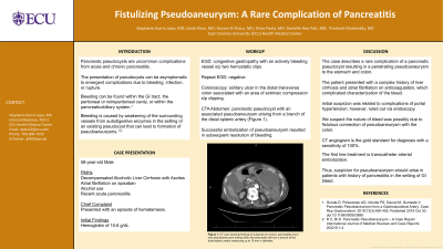
Case Description/Methods: A 58-year-old male with a history of decompensated alcoholic liver cirrhosis with ascites, atrial fibrillation on apixaban, alcohol use, and recent acute pancreatitis presented with an episode of hematemesis. The patient was anemic with a hemoglobin of 10.6 g/dL. EGD showed congestive gastropathy with an actively bleeding vessel requiring placement of two hemostatic clips resulting in temporary resolution of bleeding (Figure 1). However, he had another episode of hematemesis this time associated with hematochezia. Repeat EGD was unrevealing; therefore, a colonoscopy was done which demonstrated a solitary ulcer in the distal transverse colon associated with an area of extrinsic compression which`h was endoscopically clipped. He continued to have hematochezia and CTA demonstrated a pancreatic pseudocyst with an associated pseudoaneurysm arising from a branch of the distal splenic artery. This was promptly successfully embolized, leading to subsequent resolution of bleeding.
Discussion: The case above describes a rare complication of a pancreatic pseudocyst resulting in a penetrating pseudoaneurysm to the stomach and colon. Our patient presented with a complex history of liver cirrhosis and atrial fibrillation on anticoagulation, which complicated characterization of the bleed, as our initial suspicion was related to complications of portal hypertension; however, upon further endoscopic evaluation this was not found. Therefore, we suspect the nature of bleed was possibly due to fistulous connection of pseudoaneurysm with the colon. CT angiogram is the gold standard for diagnosis with a sensitivity of 100%. The first line treatment is transcatheter arterial embolization. Thus, suspicion for pseudoaneurysm should arise in patients with history of pancreatitis in the setting of GI bleed.

Disclosures:
Stephanie Ibarra, MD1, Zarak Khan, MD1, Shiva Poola, MD2, Husam El Sharu, MD1, Danielle Hoo-Fatt, MD1, Prashant Mudireddy, MD1. P1557 - Fistulizing Pseudoaneurysm: A Rare Complication of Pancreatitis, ACG 2023 Annual Scientific Meeting Abstracts. Vancouver, BC, Canada: American College of Gastroenterology.
1East Carolina University, Greenville, NC; 2ECU Health Medical Center, Greenville, NC
Introduction: Pancreatic pseudocysts are uncommon complications from acute and chronic pancreatitis. The presentation of pseudocysts can be asymptomatic to emergent complications due to bleeding, infection, or rupture. The development of a pseudoaneurysm are caused by weakening of the surrounding vessels from autodigestive enzymes in the setting of an existing pseudocyst. Bleeding from pseudoaneurysms can be found within the GI tract, the peritoneal or retroperitoneal cavity, or within the pancreaticobiliary system. This case highlights a patient with an obscure recurrent GI bleeding who was found to have a fistulizing pseudoaneurysm to the colon.
Case Description/Methods: A 58-year-old male with a history of decompensated alcoholic liver cirrhosis with ascites, atrial fibrillation on apixaban, alcohol use, and recent acute pancreatitis presented with an episode of hematemesis. The patient was anemic with a hemoglobin of 10.6 g/dL. EGD showed congestive gastropathy with an actively bleeding vessel requiring placement of two hemostatic clips resulting in temporary resolution of bleeding (Figure 1). However, he had another episode of hematemesis this time associated with hematochezia. Repeat EGD was unrevealing; therefore, a colonoscopy was done which demonstrated a solitary ulcer in the distal transverse colon associated with an area of extrinsic compression which`h was endoscopically clipped. He continued to have hematochezia and CTA demonstrated a pancreatic pseudocyst with an associated pseudoaneurysm arising from a branch of the distal splenic artery. This was promptly successfully embolized, leading to subsequent resolution of bleeding.
Discussion: The case above describes a rare complication of a pancreatic pseudocyst resulting in a penetrating pseudoaneurysm to the stomach and colon. Our patient presented with a complex history of liver cirrhosis and atrial fibrillation on anticoagulation, which complicated characterization of the bleed, as our initial suspicion was related to complications of portal hypertension; however, upon further endoscopic evaluation this was not found. Therefore, we suspect the nature of bleed was possibly due to fistulous connection of pseudoaneurysm with the colon. CT angiogram is the gold standard for diagnosis with a sensitivity of 100%. The first line treatment is transcatheter arterial embolization. Thus, suspicion for pseudoaneurysm should arise in patients with history of pancreatitis in the setting of GI bleed.

Figure: CT scan showing findings of subacute on chronic pancreatitis and a new pseudoaneurysm arising within the pancreatic tail from a branch of the distal splenic artery measuring up to 19 mm in diameter. There is complex surrounding pseudocyst which appears to protrude into the lumen of the splenic flexure in the left upper quadrant and hemorrhage from this pseudoaneurysm may explain lower GI bleeding.
Disclosures:
Stephanie Ibarra indicated no relevant financial relationships.
Zarak Khan indicated no relevant financial relationships.
Shiva Poola indicated no relevant financial relationships.
Husam El Sharu indicated no relevant financial relationships.
Danielle Hoo-Fatt indicated no relevant financial relationships.
Prashant Mudireddy indicated no relevant financial relationships.
Stephanie Ibarra, MD1, Zarak Khan, MD1, Shiva Poola, MD2, Husam El Sharu, MD1, Danielle Hoo-Fatt, MD1, Prashant Mudireddy, MD1. P1557 - Fistulizing Pseudoaneurysm: A Rare Complication of Pancreatitis, ACG 2023 Annual Scientific Meeting Abstracts. Vancouver, BC, Canada: American College of Gastroenterology.
