Monday Poster Session
Category: Biliary/Pancreas
P1564 - A Case of Postoperative Biliary Leak in a Patient With Duplicated Cystic Ducts
Monday, October 23, 2023
10:30 AM - 4:15 PM PT
Location: Exhibit Hall

Has Audio
- GM
Giri Movva, MD
University of Texas Medical Branch
Galveston, TX
Presenting Author(s)
Giri Movva, MD, Jordan Malone, DO, Jaison S. John, MD, Patrick Sweet, DO
University of Texas Medical Branch, Galveston, TX
Introduction: Duplicated cystic ducts with a single gallbladder is a rare congenital malformation that is typically diagnosed intraoperatively or postoperatively due to complications. It is important to identify these anomalies in the periprocedural setting to reduce the risk of postoperative complications including bile duct injuries and subsequent biliary leaks that increase postoperative morbidity and mortality.
Case Description/Methods:
A 62-year-old male with biliary dyskinesia status-post cholecystectomy was transferred from an outside hospital (OSH) to our institution with symptoms of non-radiating epigastric pain, nausea, vomiting, and chills. Interval history includes a laparoscopic cholecystectomy two months prior to presentation where duplicated cystic ducts were ligated intraoperatively at an OSH. Computed Tomography (CT) abdomen showed a bile leak with biloma formation posterior to the left lobe of the liver. Endoscopic retrograde cholangiopancreatography (ERCP) revealed papillary stenosis with common bile duct dilation but no apparent leak on the cholangiogram. A plastic biliary stent was placed, and the collection was drained. A follow-up Magnetic Resonance Imaging showed a persistent fluid collection and a Hepatobiliary iminodiacetic acid (HIDA) scan showed a persistent biliary leak into the subhepatic space. The plastic biliary stent was replaced with a metallic stent and the collection was drained with symptomatic improvement.
On presentation, lab work showed an elevated alkaline phosphatase 344 U/L. CT abdomen showed the development of a 2.9 cm fluid collection adjacent to the subhepatic drain. He underwent biliary drainage interrogation showing right-sided biliary ducts with no communication with the common bile duct. Magnetic resonance cholangiopancreatography revealed a perihepatic fluid collection with communication to the right-sided bile duct. Attempts for catheterization of the bile leak were unsuccessful with subsequent dissection of the ducts into hepatic parenchyma. An external biliary drain was placed. Right cystic duct embolization was re-attempted, but unsuccessful due to persistent duct dissection. The patient was discharged with plans for outpatient embolization.
Discussion: Treatment options for biliary leak consist of ERCP with stenting, percutaneous drainage, and duct embolization. Roux-en Y hepaticojejunostomy tends to be a last resort. It is imperative to understand post-operative complications and treatment to minimize the need for more invasive procedures.

Disclosures:
Giri Movva, MD, Jordan Malone, DO, Jaison S. John, MD, Patrick Sweet, DO. P1564 - A Case of Postoperative Biliary Leak in a Patient With Duplicated Cystic Ducts, ACG 2023 Annual Scientific Meeting Abstracts. Vancouver, BC, Canada: American College of Gastroenterology.
University of Texas Medical Branch, Galveston, TX
Introduction: Duplicated cystic ducts with a single gallbladder is a rare congenital malformation that is typically diagnosed intraoperatively or postoperatively due to complications. It is important to identify these anomalies in the periprocedural setting to reduce the risk of postoperative complications including bile duct injuries and subsequent biliary leaks that increase postoperative morbidity and mortality.
Case Description/Methods:
A 62-year-old male with biliary dyskinesia status-post cholecystectomy was transferred from an outside hospital (OSH) to our institution with symptoms of non-radiating epigastric pain, nausea, vomiting, and chills. Interval history includes a laparoscopic cholecystectomy two months prior to presentation where duplicated cystic ducts were ligated intraoperatively at an OSH. Computed Tomography (CT) abdomen showed a bile leak with biloma formation posterior to the left lobe of the liver. Endoscopic retrograde cholangiopancreatography (ERCP) revealed papillary stenosis with common bile duct dilation but no apparent leak on the cholangiogram. A plastic biliary stent was placed, and the collection was drained. A follow-up Magnetic Resonance Imaging showed a persistent fluid collection and a Hepatobiliary iminodiacetic acid (HIDA) scan showed a persistent biliary leak into the subhepatic space. The plastic biliary stent was replaced with a metallic stent and the collection was drained with symptomatic improvement.
On presentation, lab work showed an elevated alkaline phosphatase 344 U/L. CT abdomen showed the development of a 2.9 cm fluid collection adjacent to the subhepatic drain. He underwent biliary drainage interrogation showing right-sided biliary ducts with no communication with the common bile duct. Magnetic resonance cholangiopancreatography revealed a perihepatic fluid collection with communication to the right-sided bile duct. Attempts for catheterization of the bile leak were unsuccessful with subsequent dissection of the ducts into hepatic parenchyma. An external biliary drain was placed. Right cystic duct embolization was re-attempted, but unsuccessful due to persistent duct dissection. The patient was discharged with plans for outpatient embolization.
Discussion: Treatment options for biliary leak consist of ERCP with stenting, percutaneous drainage, and duct embolization. Roux-en Y hepaticojejunostomy tends to be a last resort. It is imperative to understand post-operative complications and treatment to minimize the need for more invasive procedures.

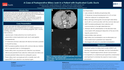
Figure: Figure 1: CT Abdomen Pelvis with contrast axial view - 2.9 cm fluid collection near the right subhepatic region.
Figure 2: MRCP-right sided bile duct communication with perihepatic fluid collection.
Figure 2: MRCP-right sided bile duct communication with perihepatic fluid collection.
Disclosures:
Giri Movva indicated no relevant financial relationships.
Jordan Malone indicated no relevant financial relationships.
Jaison John indicated no relevant financial relationships.
Patrick Sweet indicated no relevant financial relationships.
Giri Movva, MD, Jordan Malone, DO, Jaison S. John, MD, Patrick Sweet, DO. P1564 - A Case of Postoperative Biliary Leak in a Patient With Duplicated Cystic Ducts, ACG 2023 Annual Scientific Meeting Abstracts. Vancouver, BC, Canada: American College of Gastroenterology.
