Monday Poster Session
Category: Biliary/Pancreas
P1566 - Rare Primary Pancreatic Squamous Cell Carcinoma Presenting as Duodenal Obstruction
Monday, October 23, 2023
10:30 AM - 4:15 PM PT
Location: Exhibit Hall

Has Audio

Analine B. Delgado, BS
Advocate Lutheran General Hospital
Chicago, IL
Presenting Author(s)
Analine Delgado, BS1, Carey August, MD2, Faris M. Murad, MD3
1Advocate Lutheran General Hospital, Chicago, IL; 2Advocate Illinois Masonic Medical Center, Chicago, IL; 3Advocate Lutheran General Hospital, Park Ridge, IL
Introduction: Squamous cell carcinoma (SCC) is a malignant tumor that usually originates from the squamous epithelium of various organs, such as the skin, digestive and respiratory tracts. Primary SCC of the pancreas (SCCP) is exceedingly rare, accounting for approximately 1% of all pancreatic malignancies, with a reported incidence rate of 0.5-5%. This rarity makes SCCP a diagnostic and therapeutic challenge, as its presentation and imaging features are not well established. Given that squamous cells do not normally exist in the parenchyma of the pancreas, extensive workup is necessary to exclude other primary metastatic sites. We describe a rare case of primary SCCP presenting as duodenal obstruction.
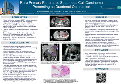
Case Description/Methods: A 56-year-old female presented with progressive symptoms of gastric outlet obstruction, nausea, vomiting, and unintentional weight loss. Upper endoscopy revealed an acquired extrinsic severe stenosis of the third portion of the duodenum that was non-traversed, and biopsy specimens from the affected area showed no evidence of malignancy. CT scan of the abdomen and pelvis revealed a suspected solid pancreatic neoplasm. Further evaluation with an upper endoscopic ultrasound (EUS) revealed an irregular mass in the pancreatic tail, staged as T4 N1 Mx by endosonographic criteria. The mass was hypoechoic, and sonographic evidence suggested encasement of the superior mesenteric artery, the celiac trunk, and the splenic artery. Tissue acquisition during EUS was obtained using a 22G core biopsy needle. Pathology of the pancreatic mass specimen revealed pure squamous cell differentiation. Due to the advanced stage of the disease and vascular involvement, the patient was not a surgical candidate. She was started on palliative chemotherapy, received enteral stenting to relieve her gastric outlet obstruction, and unfortunately passed away a few months later.
Discussion: SCCP is an aggressive malignancy with a variety of symptoms, clinically presenting similarly to that of ductal adenocarcinoma. Due to its rarity, SCCP can be difficult to diagnose, and patients may not receive a definitive diagnosis until advanced stages of the disease. In our case, EUS with fine needle core biopsy was important as it provided enough intact tissue for adequate histopathological analysis to make the diagnosis. SCCP carries a dismal prognosis, with a median survival of < 6 months, and limited treatment options highlight the importance of early recognition and intervention.

Disclosures:
Analine Delgado, BS1, Carey August, MD2, Faris M. Murad, MD3. P1566 - Rare Primary Pancreatic Squamous Cell Carcinoma Presenting as Duodenal Obstruction, ACG 2023 Annual Scientific Meeting Abstracts. Vancouver, BC, Canada: American College of Gastroenterology.
1Advocate Lutheran General Hospital, Chicago, IL; 2Advocate Illinois Masonic Medical Center, Chicago, IL; 3Advocate Lutheran General Hospital, Park Ridge, IL
Introduction: Squamous cell carcinoma (SCC) is a malignant tumor that usually originates from the squamous epithelium of various organs, such as the skin, digestive and respiratory tracts. Primary SCC of the pancreas (SCCP) is exceedingly rare, accounting for approximately 1% of all pancreatic malignancies, with a reported incidence rate of 0.5-5%. This rarity makes SCCP a diagnostic and therapeutic challenge, as its presentation and imaging features are not well established. Given that squamous cells do not normally exist in the parenchyma of the pancreas, extensive workup is necessary to exclude other primary metastatic sites. We describe a rare case of primary SCCP presenting as duodenal obstruction.
Case Description/Methods: A 56-year-old female presented with progressive symptoms of gastric outlet obstruction, nausea, vomiting, and unintentional weight loss. Upper endoscopy revealed an acquired extrinsic severe stenosis of the third portion of the duodenum that was non-traversed, and biopsy specimens from the affected area showed no evidence of malignancy. CT scan of the abdomen and pelvis revealed a suspected solid pancreatic neoplasm. Further evaluation with an upper endoscopic ultrasound (EUS) revealed an irregular mass in the pancreatic tail, staged as T4 N1 Mx by endosonographic criteria. The mass was hypoechoic, and sonographic evidence suggested encasement of the superior mesenteric artery, the celiac trunk, and the splenic artery. Tissue acquisition during EUS was obtained using a 22G core biopsy needle. Pathology of the pancreatic mass specimen revealed pure squamous cell differentiation. Due to the advanced stage of the disease and vascular involvement, the patient was not a surgical candidate. She was started on palliative chemotherapy, received enteral stenting to relieve her gastric outlet obstruction, and unfortunately passed away a few months later.
Discussion: SCCP is an aggressive malignancy with a variety of symptoms, clinically presenting similarly to that of ductal adenocarcinoma. Due to its rarity, SCCP can be difficult to diagnose, and patients may not receive a definitive diagnosis until advanced stages of the disease. In our case, EUS with fine needle core biopsy was important as it provided enough intact tissue for adequate histopathological analysis to make the diagnosis. SCCP carries a dismal prognosis, with a median survival of < 6 months, and limited treatment options highlight the importance of early recognition and intervention.

Figure: Figure 1.
A: Endoscopic ultrasound (EUS) showing an irregular, hypoechoic mass in the pancreatic tail that was biopsied using Medtronic SharkCore Biopsy needle (arrowhead).
Pancreatic Biopsy Specimen. All images are hematoxylin and eosin stained (H&E).
B: (Original magnification X 40) necrotic debris with nests of tumor cells;
C: (Original magnification X 100) nests of tumor cells within a desmoplastic stroma;
D: (Original magnification X 400) tumor cells showing eosinophilic cytoplasm, plate-like growth, and intercellular bridges characteristic of squamous cell carcinoma. The pleomorphic nuclei clearly indicate malignancy. Taken together, these findings are consistent with a diagnosis of squamous cell carcinoma of the pancreas.
Disclosures:
Analine Delgado indicated no relevant financial relationships.
Carey August indicated no relevant financial relationships.
Faris Murad indicated no relevant financial relationships.
Analine Delgado, BS1, Carey August, MD2, Faris M. Murad, MD3. P1566 - Rare Primary Pancreatic Squamous Cell Carcinoma Presenting as Duodenal Obstruction, ACG 2023 Annual Scientific Meeting Abstracts. Vancouver, BC, Canada: American College of Gastroenterology.

