Monday Poster Session
Category: Biliary/Pancreas
P1570 - Type 4 Choledochal Cyst: Biliary Dilation With Concomitant Stricture Presenting a Diagnostic and Treatment Challenge
Monday, October 23, 2023
10:30 AM - 4:15 PM PT
Location: Exhibit Hall

Has Audio

Sarah Grebennikov, DO, MHS
Riverside Methodist Hospital
Columbus, OH
Presenting Author(s)
Sarah Grebennikov, DO, MHS1, Patrick Salibi, MD1, Melinda Rogers, MD2, David Lo, MD, FACG2
1Riverside Methodist Hospital, Columbus, OH; 2Riverside Methodist Hospital/Ohio Gastroenterology Group, Inc., Columbus, OH
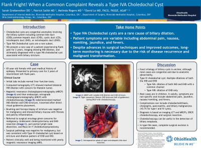
Introduction: Type lV choledochal cyst is a rare congenital malformation of the biliary tree. Type IVA have cystic dilation of both the intrahepatic (IHD) and extrahepatic bile ducts (EHD), while type IVB involves the IHD alone. Patient symptoms can vary from asymptomatic to abdominal pain, nausea, vomiting, jaundice, and fevers. We present a rare case of a patient found to have incidental image findings of IHD dilation ultimately diagnosed with a type lVA choledochal cyst associated with biliary stricture.
Case Description/Methods: 65-year-old female with a history of epilepsy initially presented to her primary care doctor’s office for intermittent left flank pain. CT followed by MRI were remarkable for marked IHD dilation with abrupt transition at the common hepatic duct (CHD), without an obvious mass (A). There was no evidence of distal common bile duct dilation. Liver function tests were normal. She was then referred for gastroenterology evaluation. EUS was negative for a mass or lymphadenopathy. ERCP with choledochoscopy noted a tight concentric fibrotic stricture at the CHD (B). Brush cytology and choledochoscopy mini forceps biopsy of the stricture were negative for malignant cells and revealed only fibrosis with patchy inflammation. A 10 french biliary stent was placed. The patient was referred to surgical oncology given concern for occult malignancy. She underwent diagnostic laparoscopy with cholecystectomy and extrahepatic bile duct resection (C), peri-portal lymph node dissection, and Roux-en-Y choledochojejunostomy. Final pathology was negative for malignancy, and demonstrated foci of stricture and dilation of the EHD and IHD consistent with a type IVA choledochal cyst.
Discussion: Type lV choledochal cysts are a rare cause of biliary duct dilation. They are associated with a high risk of complications, such as obstructive jaundice, cholangitis, and biliary malignancies. Diagnosis is typically made through cross sectional imaging. Choledochoscopy can be utilized to evaluate for dysplasia in a biliary cyst. Surgical intervention is the mainstay of treatment, with complete excision of the extrahepatic portion of the cyst with bilio-enteric anastomosis. Despite advances in surgical techniques and improved outcomes, long-term monitoring is necessary due to the risk of disease recurrence and malignant transformation.

Disclosures:
Sarah Grebennikov, DO, MHS1, Patrick Salibi, MD1, Melinda Rogers, MD2, David Lo, MD, FACG2. P1570 - Type 4 Choledochal Cyst: Biliary Dilation With Concomitant Stricture Presenting a Diagnostic and Treatment Challenge, ACG 2023 Annual Scientific Meeting Abstracts. Vancouver, BC, Canada: American College of Gastroenterology.
1Riverside Methodist Hospital, Columbus, OH; 2Riverside Methodist Hospital/Ohio Gastroenterology Group, Inc., Columbus, OH
Introduction: Type lV choledochal cyst is a rare congenital malformation of the biliary tree. Type IVA have cystic dilation of both the intrahepatic (IHD) and extrahepatic bile ducts (EHD), while type IVB involves the IHD alone. Patient symptoms can vary from asymptomatic to abdominal pain, nausea, vomiting, jaundice, and fevers. We present a rare case of a patient found to have incidental image findings of IHD dilation ultimately diagnosed with a type lVA choledochal cyst associated with biliary stricture.
Case Description/Methods: 65-year-old female with a history of epilepsy initially presented to her primary care doctor’s office for intermittent left flank pain. CT followed by MRI were remarkable for marked IHD dilation with abrupt transition at the common hepatic duct (CHD), without an obvious mass (A). There was no evidence of distal common bile duct dilation. Liver function tests were normal. She was then referred for gastroenterology evaluation. EUS was negative for a mass or lymphadenopathy. ERCP with choledochoscopy noted a tight concentric fibrotic stricture at the CHD (B). Brush cytology and choledochoscopy mini forceps biopsy of the stricture were negative for malignant cells and revealed only fibrosis with patchy inflammation. A 10 french biliary stent was placed. The patient was referred to surgical oncology given concern for occult malignancy. She underwent diagnostic laparoscopy with cholecystectomy and extrahepatic bile duct resection (C), peri-portal lymph node dissection, and Roux-en-Y choledochojejunostomy. Final pathology was negative for malignancy, and demonstrated foci of stricture and dilation of the EHD and IHD consistent with a type IVA choledochal cyst.
Discussion: Type lV choledochal cysts are a rare cause of biliary duct dilation. They are associated with a high risk of complications, such as obstructive jaundice, cholangitis, and biliary malignancies. Diagnosis is typically made through cross sectional imaging. Choledochoscopy can be utilized to evaluate for dysplasia in a biliary cyst. Surgical intervention is the mainstay of treatment, with complete excision of the extrahepatic portion of the cyst with bilio-enteric anastomosis. Despite advances in surgical techniques and improved outcomes, long-term monitoring is necessary due to the risk of disease recurrence and malignant transformation.

Figure: A) MRI Abdomen with contrast demonstrating severe dilation of main hepatic ducts and moderate intrahepatic biliary duct dilation. B) Image taken during ERCP showing concentric fibrotic stricture at the common hepatic duct. C) Diagnostic laparoscopy, cholecystectomy, and extrahepatic bile duct resection.
Disclosures:
Sarah Grebennikov indicated no relevant financial relationships.
Patrick Salibi indicated no relevant financial relationships.
Melinda Rogers indicated no relevant financial relationships.
David Lo indicated no relevant financial relationships.
Sarah Grebennikov, DO, MHS1, Patrick Salibi, MD1, Melinda Rogers, MD2, David Lo, MD, FACG2. P1570 - Type 4 Choledochal Cyst: Biliary Dilation With Concomitant Stricture Presenting a Diagnostic and Treatment Challenge, ACG 2023 Annual Scientific Meeting Abstracts. Vancouver, BC, Canada: American College of Gastroenterology.
