Monday Poster Session
Category: Colon
P1630 - A Case of Coil Assisted Retrograde Transvenous Obliteration Used to Treat Colonic Varices
Monday, October 23, 2023
10:30 AM - 4:15 PM PT
Location: Exhibit Hall

Has Audio
- MZ
Mir Zulqarnain, DO
University of Missouri-Kansas City
Kansas City, MO
Presenting Author(s)
Noor Hassan, MD1, Mir Zulqarnain, DO1, Abbas Bader, 2, Mohamed Ahmed, MD3, Travis Brown, DO1, Kavita Jadhav, MD1, Hassan Ghoz, MD1
1University of Missouri-Kansas City, Kansas City, MO; 2University of Missouri Kansas City, Kansas City, MO; 3University of Missouri Kansas City, Overland Park, KS
Introduction: Colonic variceal bleeding is a rare cause of lower GI bleeding which carries a high mortality rate. Due to limited data, optimal management of colonic variceal bleeding is unknown. Coil assisted retrograde transvenous obliteration (CARTO) has been shown to be effective in managing non-esophageal variceal bleeding, but only a few cases demonstrate its effectiveness in treating colonic variceal bleeding. We report a case of colonic varices treated with CARTO.
Case Description/Methods: A 54-year-old female with alcoholic liver cirrhosis complicated by ascites, encephalopathy, and esophageal varices was admitted with acute anemia (hemoglobin 6.5 g/dL from baseline 8 g/dL) and melena. Esophagogastroduodenoscopy (EGD) demonstrated non-bleeding Grade 1 esophageal varices. Melena resolved without intervention, but days later, the patient developed large volume hematochezia with hypotension requiring transfer to intensive care. After resuscitation, push enteroscopy and colonoscopy were performed and showed fresh blood in the ascending colon and cecum, and ascending colonic varices without evidence of active bleeding. The patient continued to have hematochezia, but computed tomography angiography of the abdomen/pelvis and RBC scintigraphy could not localize the bleeding source. Transplant hepatology did not recommend transjugular intrahepatic portosystemic shunt (TIPS) given the high MELD score of 23, acute encephalopathy, and lack of candidacy for liver transplantation. After a multi-disciplinary discussion, the decision was made to proceed with empiric eradication of the colonic varices, which were felt to be the primary bleeding source. IR performed venography and CARTO of the ascending colonic varices using a portosystemic shunt extending from the right gonadal vein which connected to the varices. Proximal splenic artery embolization was also done to decrease overall portal pressures. No persistent colonic varices were noted post-intervention and the patient had no further episodes of bleeding.
Discussion: Colonic varices pose a challenge for clinicians, as they are oftentimes difficult to identify on endoscopy or imaging, and no standardized management has been identified given the small cohort of patients. There are only a few documented cases of CRTO or modified versions of the procedure in the treatment of colonic varices. Our case, in conjunction with published data, serves as evidence that CARTO may be a useful alternative to TIPS or other invasive procedures in patients with colonic varices.

Disclosures:
Noor Hassan, MD1, Mir Zulqarnain, DO1, Abbas Bader, 2, Mohamed Ahmed, MD3, Travis Brown, DO1, Kavita Jadhav, MD1, Hassan Ghoz, MD1. P1630 - A Case of Coil Assisted Retrograde Transvenous Obliteration Used to Treat Colonic Varices, ACG 2023 Annual Scientific Meeting Abstracts. Vancouver, BC, Canada: American College of Gastroenterology.
1University of Missouri-Kansas City, Kansas City, MO; 2University of Missouri Kansas City, Kansas City, MO; 3University of Missouri Kansas City, Overland Park, KS
Introduction: Colonic variceal bleeding is a rare cause of lower GI bleeding which carries a high mortality rate. Due to limited data, optimal management of colonic variceal bleeding is unknown. Coil assisted retrograde transvenous obliteration (CARTO) has been shown to be effective in managing non-esophageal variceal bleeding, but only a few cases demonstrate its effectiveness in treating colonic variceal bleeding. We report a case of colonic varices treated with CARTO.
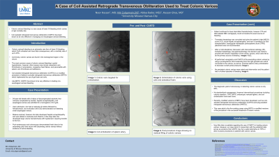
Case Description/Methods: A 54-year-old female with alcoholic liver cirrhosis complicated by ascites, encephalopathy, and esophageal varices was admitted with acute anemia (hemoglobin 6.5 g/dL from baseline 8 g/dL) and melena. Esophagogastroduodenoscopy (EGD) demonstrated non-bleeding Grade 1 esophageal varices. Melena resolved without intervention, but days later, the patient developed large volume hematochezia with hypotension requiring transfer to intensive care. After resuscitation, push enteroscopy and colonoscopy were performed and showed fresh blood in the ascending colon and cecum, and ascending colonic varices without evidence of active bleeding. The patient continued to have hematochezia, but computed tomography angiography of the abdomen/pelvis and RBC scintigraphy could not localize the bleeding source. Transplant hepatology did not recommend transjugular intrahepatic portosystemic shunt (TIPS) given the high MELD score of 23, acute encephalopathy, and lack of candidacy for liver transplantation. After a multi-disciplinary discussion, the decision was made to proceed with empiric eradication of the colonic varices, which were felt to be the primary bleeding source. IR performed venography and CARTO of the ascending colonic varices using a portosystemic shunt extending from the right gonadal vein which connected to the varices. Proximal splenic artery embolization was also done to decrease overall portal pressures. No persistent colonic varices were noted post-intervention and the patient had no further episodes of bleeding.
Discussion: Colonic varices pose a challenge for clinicians, as they are oftentimes difficult to identify on endoscopy or imaging, and no standardized management has been identified given the small cohort of patients. There are only a few documented cases of CRTO or modified versions of the procedure in the treatment of colonic varices. Our case, in conjunction with published data, serves as evidence that CARTO may be a useful alternative to TIPS or other invasive procedures in patients with colonic varices.

Figure: Images from IR procedure showing colonic varix targeted for embolization (A), embolization of colonic varix using coils and sotradecol foam (B), and post-procedure image showing no residual filling of colonic varices (C).
Disclosures:
Noor Hassan indicated no relevant financial relationships.
Mir Zulqarnain indicated no relevant financial relationships.
Abbas Bader indicated no relevant financial relationships.
Mohamed Ahmed indicated no relevant financial relationships.
Travis Brown indicated no relevant financial relationships.
Kavita Jadhav indicated no relevant financial relationships.
Hassan Ghoz indicated no relevant financial relationships.
Noor Hassan, MD1, Mir Zulqarnain, DO1, Abbas Bader, 2, Mohamed Ahmed, MD3, Travis Brown, DO1, Kavita Jadhav, MD1, Hassan Ghoz, MD1. P1630 - A Case of Coil Assisted Retrograde Transvenous Obliteration Used to Treat Colonic Varices, ACG 2023 Annual Scientific Meeting Abstracts. Vancouver, BC, Canada: American College of Gastroenterology.
