Monday Poster Session
Category: Colon
P1655 - Where Is It From? Diagnosing a Cancer of Unknown Primary via Colonoscopy
Monday, October 23, 2023
10:30 AM - 4:15 PM PT
Location: Exhibit Hall

Has Audio

Ricardo L. López-Valle, MD
VA Caribbean Healthcare System
San Juan, PR
Presenting Author(s)
Ricardo L.. López-Valle, MD, Esteban Grovas-Cordovi, MD, Kiara C.. Ortiz-Camacho, MD, José Martin-Ortiz, MD, FACG
VA Caribbean Healthcare System, San Juan, Puerto Rico
Introduction: Colorectal adenocarcinoma is one the most common cancers in the USA and worldwide. This malignancy usually arises from precursor lesions, as could be the case of a tubular adenoma with high grade dysplasia. However, it is infrequent for one to find a common pre-malignant lesion such as a tubular adenoma to be admixed with non-colonic neoplastic tissue. In the case below we relate the case of how an incidental imaging of the colon led to an unusual histologic discovery from an otherwise relatively common endoscopic finding.
Case Description/Methods: Case of a 63-year-old male with a history of liver cirrhosis secondary to alcohol use who was found with rapid progression of an apparent malignancy during a surveillance MRI. Specifically, abnormal imaging revealed new bilateral adrenal masses with a retroperitoneal soft tissue mass, suggestive of metastatic disease, and new focal wall thickening at the ascending colon, which was worrisome for an underlying primary colonic neoplasm. Of note, said findings were absent on images 4 months prior and patient had a complete colonoscopy 5 years prior without concerning pathologies. A diagnostic colonoscopy was performed in response to said abnormal imaging. Colonoscopy was remarkable for 9 large polyps. Interestingly, 2 of these 9 polyps were positive for tubular adenomas with extensive lamina propria infiltration by a poorly differentiated neoplasm that was not suggestive of colon neoplasia. Immunostains were done and negative for markers that could suggest a prostatic, melanotic, hepatocellular, lymphocytic, squamous, or neuroendocrine etiology. PET/CT later revealed multiple hypermetabolic lesions, including the left lung, bilateral adrenal glands, and the retroperitoneum. Given these findings of an aggressive metastatic disease from an unknown primary site, a goals of care conversation ensued with patient, and he opted for hospice & palliative care.
Discussion: Although a cancer from an unknown primary site is a relatively common clinical scenario, 20-25% of these cancers are poorly differentiated and cannot be recognized via routine histologic exams. Although histologic analysis was inconclusive, the poorly differentiated neoplasm found in two of the polyps did not appear to be a primary colon neoplasm. This suggests that a metastatic cancer was infiltrating the colon polyps. This showcases the importance of endoscopic sampling or resection of all identified polyps, as even seemingly benign polyps could be hiding a more life-threatening condition.

Disclosures:
Ricardo L.. López-Valle, MD, Esteban Grovas-Cordovi, MD, Kiara C.. Ortiz-Camacho, MD, José Martin-Ortiz, MD, FACG. P1655 - Where Is It From? Diagnosing a Cancer of Unknown Primary via Colonoscopy, ACG 2023 Annual Scientific Meeting Abstracts. Vancouver, BC, Canada: American College of Gastroenterology.
VA Caribbean Healthcare System, San Juan, Puerto Rico
Introduction: Colorectal adenocarcinoma is one the most common cancers in the USA and worldwide. This malignancy usually arises from precursor lesions, as could be the case of a tubular adenoma with high grade dysplasia. However, it is infrequent for one to find a common pre-malignant lesion such as a tubular adenoma to be admixed with non-colonic neoplastic tissue. In the case below we relate the case of how an incidental imaging of the colon led to an unusual histologic discovery from an otherwise relatively common endoscopic finding.
Case Description/Methods: Case of a 63-year-old male with a history of liver cirrhosis secondary to alcohol use who was found with rapid progression of an apparent malignancy during a surveillance MRI. Specifically, abnormal imaging revealed new bilateral adrenal masses with a retroperitoneal soft tissue mass, suggestive of metastatic disease, and new focal wall thickening at the ascending colon, which was worrisome for an underlying primary colonic neoplasm. Of note, said findings were absent on images 4 months prior and patient had a complete colonoscopy 5 years prior without concerning pathologies. A diagnostic colonoscopy was performed in response to said abnormal imaging. Colonoscopy was remarkable for 9 large polyps. Interestingly, 2 of these 9 polyps were positive for tubular adenomas with extensive lamina propria infiltration by a poorly differentiated neoplasm that was not suggestive of colon neoplasia. Immunostains were done and negative for markers that could suggest a prostatic, melanotic, hepatocellular, lymphocytic, squamous, or neuroendocrine etiology. PET/CT later revealed multiple hypermetabolic lesions, including the left lung, bilateral adrenal glands, and the retroperitoneum. Given these findings of an aggressive metastatic disease from an unknown primary site, a goals of care conversation ensued with patient, and he opted for hospice & palliative care.
Discussion: Although a cancer from an unknown primary site is a relatively common clinical scenario, 20-25% of these cancers are poorly differentiated and cannot be recognized via routine histologic exams. Although histologic analysis was inconclusive, the poorly differentiated neoplasm found in two of the polyps did not appear to be a primary colon neoplasm. This suggests that a metastatic cancer was infiltrating the colon polyps. This showcases the importance of endoscopic sampling or resection of all identified polyps, as even seemingly benign polyps could be hiding a more life-threatening condition.

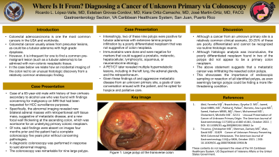
Figure: Large Polyp Transverse Colon
Disclosures:
Ricardo López-Valle indicated no relevant financial relationships.
Esteban Grovas-Cordovi indicated no relevant financial relationships.
Kiara Ortiz-Camacho indicated no relevant financial relationships.
José Martin-Ortiz indicated no relevant financial relationships.
Ricardo L.. López-Valle, MD, Esteban Grovas-Cordovi, MD, Kiara C.. Ortiz-Camacho, MD, José Martin-Ortiz, MD, FACG. P1655 - Where Is It From? Diagnosing a Cancer of Unknown Primary via Colonoscopy, ACG 2023 Annual Scientific Meeting Abstracts. Vancouver, BC, Canada: American College of Gastroenterology.
