Monday Poster Session
Category: Colon
P1661 - Incidental Discovery of Small Lymphocytic Lymphoma in a Colon Polyp
Monday, October 23, 2023
10:30 AM - 4:15 PM PT
Location: Exhibit Hall

Has Audio

Shyam Patel, MD
California Pacific Medical Center
San Francisco, CA
Presenting Author(s)
Shyam Patel, MD1, Kwun Wah Wen, MD, PhD2, Aparajita Singh, MD, MPH2
1California Pacific Medical Center, San Francisco, CA; 2University of California, San Francisco, San Francisco, CA
Introduction: Small lymphocytic lymphoma (SLL) and chronic lymphocytic leukemia (CLL) are the same disease process, described as an indolent B-cell neoplasm. SLL typically presents in the lymph nodes while CLL is found in the blood and bone marrow. Both conditions characteristically affect older adults with a median age of diagnosis at 70. Lymphadenopathy, splenomegaly, and hepatomegaly are common symptoms and signs of CLL/SLL. Gastrointestinal involvement, however, is extremely rare. We present a case of an otherwise healthy woman found to have a benign appearing cecal polyp on screening colonoscopy, which turned out to be SLL on pathology.
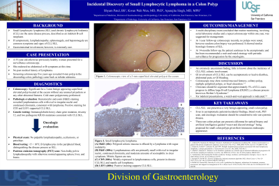
Case Description/Methods: A 55-year-old otherwise previously healthy woman presented for a screening colonoscopy. Colonoscopy was significant for a 4-mm superficial elevated polyp located at the cecum. The polyp did not have any unusual appearance. A cold snare polypectomy was performed and sent for pathologic evaluation. Hematoxylin and eosin (H&E) staining revealed lymphomatous cells with oval to irregular nuclei and condensed chromatin, consistent with lymphoma. Positive staining for CD5 and lymphoid enhancer-binding factor-1 (LEF1) supported CLL/SLL. Further gene testing revealed an immunoglobulin heavy chain gene mutation, trisomy 12, and two pathogenic KRAS mutations consistent with CLL/SLL. Since there were less than 5 × 10⁹/L B lymphocytes in the peripheral blood, the disease process was distinguished as SLL. The patient was completely asymptomatic, but we referred her for oncologic evaluation to rule out any systemic disease. Subsequent positron emission tomography (PET) scan showed non-bulky pelvic lymphadenopathy with otherwise normal appearing spleen, liver, and bones. Given the indolent and asymptomatic nature of this presentation, routine monitoring, involving serial laboratory studies and a repeat colonoscopy within one year, was suggested for management. One year out, the patient continues to be symptom-free and is awaiting a repeat colonoscopy.
Discussion: We present this case to highlight a rare diagnosis of SLL/CLL in an endoscopically benign-appearing small colon polyp. Diagnosis requires pathologic evaluation of tissue with special stains. Even in asymptomatic patients, blood work, PET scan, and oncologic evaluation should be considered to rule out systemic disease. In the absence of lymphocytosis, cytopenia, B-symptoms, or lymphadenopathy, active observation is the appropriate management approach.

Disclosures:
Shyam Patel, MD1, Kwun Wah Wen, MD, PhD2, Aparajita Singh, MD, MPH2. P1661 - Incidental Discovery of Small Lymphocytic Lymphoma in a Colon Polyp, ACG 2023 Annual Scientific Meeting Abstracts. Vancouver, BC, Canada: American College of Gastroenterology.
1California Pacific Medical Center, San Francisco, CA; 2University of California, San Francisco, San Francisco, CA
Introduction: Small lymphocytic lymphoma (SLL) and chronic lymphocytic leukemia (CLL) are the same disease process, described as an indolent B-cell neoplasm. SLL typically presents in the lymph nodes while CLL is found in the blood and bone marrow. Both conditions characteristically affect older adults with a median age of diagnosis at 70. Lymphadenopathy, splenomegaly, and hepatomegaly are common symptoms and signs of CLL/SLL. Gastrointestinal involvement, however, is extremely rare. We present a case of an otherwise healthy woman found to have a benign appearing cecal polyp on screening colonoscopy, which turned out to be SLL on pathology.
Case Description/Methods: A 55-year-old otherwise previously healthy woman presented for a screening colonoscopy. Colonoscopy was significant for a 4-mm superficial elevated polyp located at the cecum. The polyp did not have any unusual appearance. A cold snare polypectomy was performed and sent for pathologic evaluation. Hematoxylin and eosin (H&E) staining revealed lymphomatous cells with oval to irregular nuclei and condensed chromatin, consistent with lymphoma. Positive staining for CD5 and lymphoid enhancer-binding factor-1 (LEF1) supported CLL/SLL. Further gene testing revealed an immunoglobulin heavy chain gene mutation, trisomy 12, and two pathogenic KRAS mutations consistent with CLL/SLL. Since there were less than 5 × 10⁹/L B lymphocytes in the peripheral blood, the disease process was distinguished as SLL. The patient was completely asymptomatic, but we referred her for oncologic evaluation to rule out any systemic disease. Subsequent positron emission tomography (PET) scan showed non-bulky pelvic lymphadenopathy with otherwise normal appearing spleen, liver, and bones. Given the indolent and asymptomatic nature of this presentation, routine monitoring, involving serial laboratory studies and a repeat colonoscopy within one year, was suggested for management. One year out, the patient continues to be symptom-free and is awaiting a repeat colonoscopy.
Discussion: We present this case to highlight a rare diagnosis of SLL/CLL in an endoscopically benign-appearing small colon polyp. Diagnosis requires pathologic evaluation of tissue with special stains. Even in asymptomatic patients, blood work, PET scan, and oncologic evaluation should be considered to rule out systemic disease. In the absence of lymphocytosis, cytopenia, B-symptoms, or lymphadenopathy, active observation is the appropriate management approach.

Figure: Small lymphocytic lymphoma. (A) At this low power of H&E (40x), the polypoid colonic mucosa is extensively effaced by a lymphoma with vague nodularity. (B) At high-power magnification (400x), the lymphomatous cells are mostly small in size with oval to irregular nuclei, condensed chromatin, and moderate amounts of eosinophilic to clear cytoplasm. Mitotic figures are rare. (C) CD5 (100x) is weakly expressed in lymphomatous cells, which can be seen in chronic lymphocytic lymphoma/small lymphocytic lymphoma (CLL/SLL) and mantle cell lymphoma. (D) The lymphomatous cells are variably positive for LEF1 (400x), which supports CLL/SLL.
Disclosures:
Shyam Patel indicated no relevant financial relationships.
Kwun Wah Wen indicated no relevant financial relationships.
Aparajita Singh indicated no relevant financial relationships.
Shyam Patel, MD1, Kwun Wah Wen, MD, PhD2, Aparajita Singh, MD, MPH2. P1661 - Incidental Discovery of Small Lymphocytic Lymphoma in a Colon Polyp, ACG 2023 Annual Scientific Meeting Abstracts. Vancouver, BC, Canada: American College of Gastroenterology.
