Monday Poster Session
Category: Colon
P1679 - Rediscovering the Vitality: The Lost Art of the Digital Rectal Exam in Modern Healthcare
Monday, October 23, 2023
10:30 AM - 4:15 PM PT
Location: Exhibit Hall

Has Audio

Maya Mahmoud, MD
Saint Louis University
St. Louis, MO
Presenting Author(s)
Maya Mahmoud, MD1, Mahmoud Y. Madi, MD2, Pradeep Yarra, MD1, Islam Mohamed, MD3, Wissam Kiwan, MD1
1Saint Louis University, St. Louis, MO; 2Saint Louis University Hospital, St. Louis, MO; 3University of Missouri-Kansas City, Kansas City, MO
Introduction: Digital rectal examination (DRE) is an essential bedside tool to evaluate anorectal disorders. In the era of focused physical exams and the widespread availability of various imaging modalities, DRE has been often overlooked and underutilized. Often, training for the proper techniques of DRE is scarce, leading to inadequate knowledge of how to interpret findings of this simple exam. This report sheds light on the importance of digital rectal examination as a valuable and credible exam in gastrointestinal diseases.
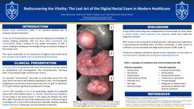
Case Description/Methods: We report the case of a 73-year-old man who was admitted for acute limb ischemia treated with endarterectomy, thrombectomy, antiplatelets agents and anticoagulants. Two-days later, the patient developed bright red blood per rectum. His hemoglobin progressively worsened during hospital stay from 15 g/dl on admission to 9.3 g/dl, without significant hemodynamic change. Careful digital rectal exam revealed a 2 to 3 cm protruding, slightly firm polypoid tissue with mild friability (Figure A). Flexible sigmoidoscopy showed a 25 mm polyp in the distal rectum 5 mm above the dentate line with NICE (NBI International Colorectal Endoscopic) class 2 features (Figure B and Figure C). The polyp was semi-pedunculated with a very thick stalk. The polyp was reducible. Biopsies revealed tubular adenoma. The patient was referred for endoscopic mucosal resection of the polyp as an outpatient.
Discussion: In our patient, DRE was able to promptly reveal the cause of lower gastrointestinal bleeding and guide the selection of the appropriate test. DRE remains a key component of physical exam, with particular importance in gastrointestinal bleeding cases, yet often forgotten. A wide variety of conditions can be evaluated and diagnosed by means of DRE (Table 1). Continuous emphasis should be placed on the importance of this exam in routine practice, but also on learning and mastering this tool.

Disclosures:
Maya Mahmoud, MD1, Mahmoud Y. Madi, MD2, Pradeep Yarra, MD1, Islam Mohamed, MD3, Wissam Kiwan, MD1. P1679 - Rediscovering the Vitality: The Lost Art of the Digital Rectal Exam in Modern Healthcare, ACG 2023 Annual Scientific Meeting Abstracts. Vancouver, BC, Canada: American College of Gastroenterology.
1Saint Louis University, St. Louis, MO; 2Saint Louis University Hospital, St. Louis, MO; 3University of Missouri-Kansas City, Kansas City, MO
Introduction: Digital rectal examination (DRE) is an essential bedside tool to evaluate anorectal disorders. In the era of focused physical exams and the widespread availability of various imaging modalities, DRE has been often overlooked and underutilized. Often, training for the proper techniques of DRE is scarce, leading to inadequate knowledge of how to interpret findings of this simple exam. This report sheds light on the importance of digital rectal examination as a valuable and credible exam in gastrointestinal diseases.
Case Description/Methods: We report the case of a 73-year-old man who was admitted for acute limb ischemia treated with endarterectomy, thrombectomy, antiplatelets agents and anticoagulants. Two-days later, the patient developed bright red blood per rectum. His hemoglobin progressively worsened during hospital stay from 15 g/dl on admission to 9.3 g/dl, without significant hemodynamic change. Careful digital rectal exam revealed a 2 to 3 cm protruding, slightly firm polypoid tissue with mild friability (Figure A). Flexible sigmoidoscopy showed a 25 mm polyp in the distal rectum 5 mm above the dentate line with NICE (NBI International Colorectal Endoscopic) class 2 features (Figure B and Figure C). The polyp was semi-pedunculated with a very thick stalk. The polyp was reducible. Biopsies revealed tubular adenoma. The patient was referred for endoscopic mucosal resection of the polyp as an outpatient.
Discussion: In our patient, DRE was able to promptly reveal the cause of lower gastrointestinal bleeding and guide the selection of the appropriate test. DRE remains a key component of physical exam, with particular importance in gastrointestinal bleeding cases, yet often forgotten. A wide variety of conditions can be evaluated and diagnosed by means of DRE (Table 1). Continuous emphasis should be placed on the importance of this exam in routine practice, but also on learning and mastering this tool.

Figure: Figure A: DRE showing prolapsed firm polypoid tissue with friability and bleeding. Figure B: Retroflexion view of the semi-pedunculated rectal polyp. Figure C: Near focus images of prolapsed rectal polyp.
Disclosures:
Maya Mahmoud indicated no relevant financial relationships.
Mahmoud Madi indicated no relevant financial relationships.
Pradeep Yarra indicated no relevant financial relationships.
Islam Mohamed indicated no relevant financial relationships.
Wissam Kiwan indicated no relevant financial relationships.
Maya Mahmoud, MD1, Mahmoud Y. Madi, MD2, Pradeep Yarra, MD1, Islam Mohamed, MD3, Wissam Kiwan, MD1. P1679 - Rediscovering the Vitality: The Lost Art of the Digital Rectal Exam in Modern Healthcare, ACG 2023 Annual Scientific Meeting Abstracts. Vancouver, BC, Canada: American College of Gastroenterology.
