Monday Poster Session
Category: Colon
P1682 - Case Report: Non-Healing Buccal Mucosa Lesion: Atypical Presentation of Metastatic Colon Adenocarcinoma
Monday, October 23, 2023
10:30 AM - 4:15 PM PT
Location: Exhibit Hall

Has Audio
- MS
Milaris Sanchez-Cordero, MD
Mayaguez Medical Center
Mayaguez, Puerto Rico
Presenting Author(s)
Milaris Sanchez-Cordero, MD1, Yue-Sai Jao-Ayala, MD2, Pablo J. Lopez-Lopez, MD1, Santa Merle-Ramirez, MD1
1Mayaguez Medical Center, Mayaguez, Puerto Rico; 2University of Puerto Rico Medical Sciences Campus, San Juan, Puerto Rico
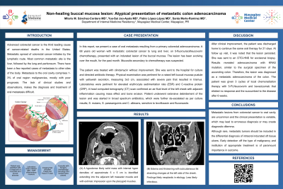
Introduction: Advanced colorectal cancer is the third leading cause of cancer-related deaths in the United States. Metastatic spread of colorectal cancer initiates by the lymphatic route. Most common metastatic site is the liver, followed by the lung and peritoneum. There have been a few reported cases of metastasis to other sites of the body. Metastasis to the oral cavity comprises 1-3% of oral region malignancies, mostly with poor prognosis. The lack of clinical studies and observations, makes the diagnosis and treatment of oral metastasis difficult. In this report, we present a case of oral metastasis resulting from a primary colorectal adenocarcinoma.
Case Description/Methods: A 68 y/o woman with metastatic colorectal cancer to lung and liver, on 5-fluorouracil/leucovorin chemotherapy, presented with an indurated lesion of the buccal mucosa. The lesion has been evolving over the mouth, for the past month. Mucositis secondary to chemotherapy was suspected. The patient was treated with clindamycin without improvement. She was sent to the hospital for culture and directed antibiotic therapy. Physical examination was pertinent for a raised left buccal mucosa pustule with yellowish secretion, measuring 3x3 cm, associated with severe pain that resulted in trismus. Lab works were pertinent for elevated ESR and CRP. A head CT scan confirmed an air-fluid level of the left cheek with adjacent inflammation causing mass effect and bone erosion. Patient underwent extensive debridement of the lesion. She was started in broad spectrum antibiotics, which were further de-escalated as per culture results; S. mutans, S. parasanguinis and C. albicans, sensitive to levofloxacin and fluconazole. After clinical improvement, the patient was discharged home to continue the same oral therapy for 21 days. At follow up visit, it was noted that the lesion persisted. She was sent to an OTO-HNS for excisional biopsy. Results revealed adenocarcinoma with KRAS mutation, similar to the surgical specimen of the ascending colon. Therefore, the lesion was diagnosed as a metastatic adenocarcinoma of the colon. The patient was given 3 cycles of local chemoradiation therapy with 5-FU/leucovorin and bevacizumab that elicited no response and the succumbed to the disease after 6 weeks.
Discussion: Although rare, metastatic tumors should be included in the differential diagnosis of intraoral indurated oft tissue ulcers. Early detection off the type of malignancy and institution of appropriate treatment is of paramount importance in outcome.
Disclosures:
Milaris Sanchez-Cordero, MD1, Yue-Sai Jao-Ayala, MD2, Pablo J. Lopez-Lopez, MD1, Santa Merle-Ramirez, MD1. P1682 - Case Report: Non-Healing Buccal Mucosa Lesion: Atypical Presentation of Metastatic Colon Adenocarcinoma, ACG 2023 Annual Scientific Meeting Abstracts. Vancouver, BC, Canada: American College of Gastroenterology.
1Mayaguez Medical Center, Mayaguez, Puerto Rico; 2University of Puerto Rico Medical Sciences Campus, San Juan, Puerto Rico
Introduction: Advanced colorectal cancer is the third leading cause of cancer-related deaths in the United States. Metastatic spread of colorectal cancer initiates by the lymphatic route. Most common metastatic site is the liver, followed by the lung and peritoneum. There have been a few reported cases of metastasis to other sites of the body. Metastasis to the oral cavity comprises 1-3% of oral region malignancies, mostly with poor prognosis. The lack of clinical studies and observations, makes the diagnosis and treatment of oral metastasis difficult. In this report, we present a case of oral metastasis resulting from a primary colorectal adenocarcinoma.
Case Description/Methods: A 68 y/o woman with metastatic colorectal cancer to lung and liver, on 5-fluorouracil/leucovorin chemotherapy, presented with an indurated lesion of the buccal mucosa. The lesion has been evolving over the mouth, for the past month. Mucositis secondary to chemotherapy was suspected. The patient was treated with clindamycin without improvement. She was sent to the hospital for culture and directed antibiotic therapy. Physical examination was pertinent for a raised left buccal mucosa pustule with yellowish secretion, measuring 3x3 cm, associated with severe pain that resulted in trismus. Lab works were pertinent for elevated ESR and CRP. A head CT scan confirmed an air-fluid level of the left cheek with adjacent inflammation causing mass effect and bone erosion. Patient underwent extensive debridement of the lesion. She was started in broad spectrum antibiotics, which were further de-escalated as per culture results; S. mutans, S. parasanguinis and C. albicans, sensitive to levofloxacin and fluconazole. After clinical improvement, the patient was discharged home to continue the same oral therapy for 21 days. At follow up visit, it was noted that the lesion persisted. She was sent to an OTO-HNS for excisional biopsy. Results revealed adenocarcinoma with KRAS mutation, similar to the surgical specimen of the ascending colon. Therefore, the lesion was diagnosed as a metastatic adenocarcinoma of the colon. The patient was given 3 cycles of local chemoradiation therapy with 5-FU/leucovorin and bevacizumab that elicited no response and the succumbed to the disease after 6 weeks.
Discussion: Although rare, metastatic tumors should be included in the differential diagnosis of intraoral indurated oft tissue ulcers. Early detection off the type of malignancy and institution of appropriate treatment is of paramount importance in outcome.
Disclosures:
Milaris Sanchez-Cordero indicated no relevant financial relationships.
Yue-Sai Jao-Ayala indicated no relevant financial relationships.
Pablo Lopez-Lopez indicated no relevant financial relationships.
Santa Merle-Ramirez indicated no relevant financial relationships.
Milaris Sanchez-Cordero, MD1, Yue-Sai Jao-Ayala, MD2, Pablo J. Lopez-Lopez, MD1, Santa Merle-Ramirez, MD1. P1682 - Case Report: Non-Healing Buccal Mucosa Lesion: Atypical Presentation of Metastatic Colon Adenocarcinoma, ACG 2023 Annual Scientific Meeting Abstracts. Vancouver, BC, Canada: American College of Gastroenterology.
