Monday Poster Session
Category: Colon
P1685 - A Rare Case of Localized Colonic Amyloidosis Identified During a Screening Colonoscopy
Monday, October 23, 2023
10:30 AM - 4:15 PM PT
Location: Exhibit Hall

Has Audio
- CA
Chmsalddin Alkhas, MD
Mansoura University
Toledo, OH
Presenting Author(s)
Chmsalddin Alkhas, MD1, Sami Ghazaleh, MD2, Ali Heif, 3, Sara Stanley, DO2, Muhannad Heif, MD4
1Mansoura University, Toledo, OH; 2University of Toledo, Toledo, OH; 3St. John’s Jesuit High School, Toledo, OH; 4University of Toledo, Promedica Healthcare System, Toledo, OH
Introduction: Amyloidosis is an abnormal accumulation of amyloid protein in different organs and tissues, which typically results in nephropathy, cardiomegaly, hepatomegaly, and neuropathy. We report a rare case of localized amyloidosis that was identified during a screening colonoscopy.
Case Description/Methods: A 73-year-old male patient was referred to the gastroenterology clinic for a screening colonoscopy. Past medical history was significant for essential hypertension, type 2 diabetes mellites, gastroesophageal reflux disease (GERD), hypothyroidism, and myasthenia gravis. Past surgical history was significant for cholecystectomy. Family history was positive for colorectal cancer in maternal uncle at the age of 45. He was a former smoker and denied alcohol or illicit drug use.
Screening colonoscopy revealed one 12 mm flat polyp in the ascending colon. The polyp was removed with endoscopic mucosal resection (EMR) and retrieved successfully. Biopsy of the polyp showed amorphous deposits in the submucosa, suggestive of amyloid deposits. Congo red stain was performed, and stain was suggestive of amyloidosis. Liquid chromatography tandem mass spectrometry was later performed on the biopsy, which detected a peptide profile consistent with AL (kappa)-type amyloid deposition. In addition, seven other sub-centimeter tubular adenomas were seen in the transverse and sigmoid colons, which were removed with cold snare and cold biopsy forceps.
The patient was referred to hematology to rule out systemic amyloidosis. Extensive workup by hematology was negative for systemic amyloidosis including absence of proteinuria, Bence-Jones proteins, monoclonal spike in serum protein immunofixation. The only abnormal finding was a slightly elevated kappa/lambda light chain ratio at 1.69. In addition, fat pad biopsy showed no evidence of Congo red/amyloid deposits, cardiac MRI showed no evidence of amyloidosis, and bone marrow biopsy showed no evidence of plasma cell dyscrasia and was negative for amyloid stain. It was concluded that the patient had localized amyloidosis without evidence of systemic disease.
Discussion: Gastrointestinal amyloidosis is a common finding in systemic amyloidosis, especially AA amyloidosis. However, localized gastrointestinal amyloidosis without evidence of systemic amyloidosis is uncommon. Management consists of observation or removal of the localized deposition. Patients have a good prognosis and they do not typically transition to systemic amyloidosis.

Disclosures:
Chmsalddin Alkhas, MD1, Sami Ghazaleh, MD2, Ali Heif, 3, Sara Stanley, DO2, Muhannad Heif, MD4. P1685 - A Rare Case of Localized Colonic Amyloidosis Identified During a Screening Colonoscopy, ACG 2023 Annual Scientific Meeting Abstracts. Vancouver, BC, Canada: American College of Gastroenterology.
1Mansoura University, Toledo, OH; 2University of Toledo, Toledo, OH; 3St. John’s Jesuit High School, Toledo, OH; 4University of Toledo, Promedica Healthcare System, Toledo, OH
Introduction: Amyloidosis is an abnormal accumulation of amyloid protein in different organs and tissues, which typically results in nephropathy, cardiomegaly, hepatomegaly, and neuropathy. We report a rare case of localized amyloidosis that was identified during a screening colonoscopy.
Case Description/Methods: A 73-year-old male patient was referred to the gastroenterology clinic for a screening colonoscopy. Past medical history was significant for essential hypertension, type 2 diabetes mellites, gastroesophageal reflux disease (GERD), hypothyroidism, and myasthenia gravis. Past surgical history was significant for cholecystectomy. Family history was positive for colorectal cancer in maternal uncle at the age of 45. He was a former smoker and denied alcohol or illicit drug use.
Screening colonoscopy revealed one 12 mm flat polyp in the ascending colon. The polyp was removed with endoscopic mucosal resection (EMR) and retrieved successfully. Biopsy of the polyp showed amorphous deposits in the submucosa, suggestive of amyloid deposits. Congo red stain was performed, and stain was suggestive of amyloidosis. Liquid chromatography tandem mass spectrometry was later performed on the biopsy, which detected a peptide profile consistent with AL (kappa)-type amyloid deposition. In addition, seven other sub-centimeter tubular adenomas were seen in the transverse and sigmoid colons, which were removed with cold snare and cold biopsy forceps.
The patient was referred to hematology to rule out systemic amyloidosis. Extensive workup by hematology was negative for systemic amyloidosis including absence of proteinuria, Bence-Jones proteins, monoclonal spike in serum protein immunofixation. The only abnormal finding was a slightly elevated kappa/lambda light chain ratio at 1.69. In addition, fat pad biopsy showed no evidence of Congo red/amyloid deposits, cardiac MRI showed no evidence of amyloidosis, and bone marrow biopsy showed no evidence of plasma cell dyscrasia and was negative for amyloid stain. It was concluded that the patient had localized amyloidosis without evidence of systemic disease.
Discussion: Gastrointestinal amyloidosis is a common finding in systemic amyloidosis, especially AA amyloidosis. However, localized gastrointestinal amyloidosis without evidence of systemic amyloidosis is uncommon. Management consists of observation or removal of the localized deposition. Patients have a good prognosis and they do not typically transition to systemic amyloidosis.

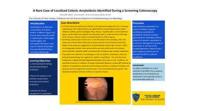
Figure: Polyp seen during colonoscopy prior to removal
Disclosures:
Chmsalddin Alkhas indicated no relevant financial relationships.
Sami Ghazaleh indicated no relevant financial relationships.
Ali Heif indicated no relevant financial relationships.
Sara Stanley indicated no relevant financial relationships.
Muhannad Heif indicated no relevant financial relationships.
Chmsalddin Alkhas, MD1, Sami Ghazaleh, MD2, Ali Heif, 3, Sara Stanley, DO2, Muhannad Heif, MD4. P1685 - A Rare Case of Localized Colonic Amyloidosis Identified During a Screening Colonoscopy, ACG 2023 Annual Scientific Meeting Abstracts. Vancouver, BC, Canada: American College of Gastroenterology.
