Monday Poster Session
Category: Colon
P1702 - Sigmoid Submucosal Hematoma: A Complication of Colonoscopy in a Healthy 51-Year-Old
Monday, October 23, 2023
10:30 AM - 4:15 PM PT
Location: Exhibit Hall

Has Audio

Sara Kamionkowski, DO
MetroHealth Medical Center
Cleveland, OH
Presenting Author(s)
Sara Kamionkowski, DO1, Shaman Dalal, MD1, Neil Patel, MD1, Michael Kurin, MD2
1MetroHealth Medical Center, Cleveland, OH; 2MetroHealth, Cleveland, OH
Introduction: Colonic submucosal hematomas are potential complications of colonoscopy, especially in patients with bleeding disorders or taking anticoagulants. Here, we present a case of colonic submucosal hematoma in an otherwise healthy patient with no risk factors for bleeding.
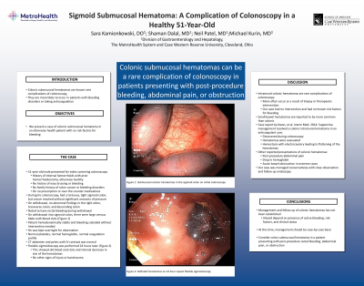
Case Description/Methods: A healthy 51-year-old male presented for his first screening colonoscopy. He had no family or personal history of colon cancer or bleeding disorders. He was not taking any medications including anticoagulants. During his colonoscopy he was noted to have a tortuous sigmoid colon with difficulty passing the scope. The cecum was able to be reached with no abnormal findings on withdraw through the descending colon. During his procedure, he was noted to have hematochezia. Upon withdrawal of the scope in the sigmoid colon, there were large venous blebs noted with blood with clots (Figure 1a). Bleeding subsided after washing. He remained stable, however given the extent of bleeding, the patient was observed overnight. Lab tests showed normal platelets, hemoglobin, and coagulation profile. CT abdomen and pelvis with intravenous contrast did not show any perforation or signs of further bleeding. A flexible sigmoidoscopy was performed the following day to assess for any mucosal injury. The two submucosal hematomas in the sigmoid colon had decreased in size and likely burst (Figure 1b).
Discussion: Colonic hematomas are rare complications of colonoscopy usually because of biopsy or therapeutic intervention, especially in patients taking anticoagulants or with bleeding disorders. It is unclear what risk factors would have led to the hematomas in the case we describe. Small bowel hematomas have been reported more frequently than colonic. There are few case reports of colonic intramural hematomas, each with different causes and treatment. One case report described colonic hematomas found during colonoscopy which were evacuated and treated with electrocautery. Other reports describe delayed presentation with abdominal pain and signs of bleeding. Some extreme cases have presented as bowel obstruction. Management and follow up of these hematomas has not been established but depends on presence of active bleeding and clinical status. Our case was managed with close follow up endoscopy to ensure resolution of the hematomas. Colonic intramural hematomas should be considered in patients with brisk lower gastrointestinal bleeding following colonoscopy or in patients with post-procedure abdominal pain or obstruction.

Disclosures:
Sara Kamionkowski, DO1, Shaman Dalal, MD1, Neil Patel, MD1, Michael Kurin, MD2. P1702 - Sigmoid Submucosal Hematoma: A Complication of Colonoscopy in a Healthy 51-Year-Old, ACG 2023 Annual Scientific Meeting Abstracts. Vancouver, BC, Canada: American College of Gastroenterology.
1MetroHealth Medical Center, Cleveland, OH; 2MetroHealth, Cleveland, OH
Introduction: Colonic submucosal hematomas are potential complications of colonoscopy, especially in patients with bleeding disorders or taking anticoagulants. Here, we present a case of colonic submucosal hematoma in an otherwise healthy patient with no risk factors for bleeding.
Case Description/Methods: A healthy 51-year-old male presented for his first screening colonoscopy. He had no family or personal history of colon cancer or bleeding disorders. He was not taking any medications including anticoagulants. During his colonoscopy he was noted to have a tortuous sigmoid colon with difficulty passing the scope. The cecum was able to be reached with no abnormal findings on withdraw through the descending colon. During his procedure, he was noted to have hematochezia. Upon withdrawal of the scope in the sigmoid colon, there were large venous blebs noted with blood with clots (Figure 1a). Bleeding subsided after washing. He remained stable, however given the extent of bleeding, the patient was observed overnight. Lab tests showed normal platelets, hemoglobin, and coagulation profile. CT abdomen and pelvis with intravenous contrast did not show any perforation or signs of further bleeding. A flexible sigmoidoscopy was performed the following day to assess for any mucosal injury. The two submucosal hematomas in the sigmoid colon had decreased in size and likely burst (Figure 1b).
Discussion: Colonic hematomas are rare complications of colonoscopy usually because of biopsy or therapeutic intervention, especially in patients taking anticoagulants or with bleeding disorders. It is unclear what risk factors would have led to the hematomas in the case we describe. Small bowel hematomas have been reported more frequently than colonic. There are few case reports of colonic intramural hematomas, each with different causes and treatment. One case report described colonic hematomas found during colonoscopy which were evacuated and treated with electrocautery. Other reports describe delayed presentation with abdominal pain and signs of bleeding. Some extreme cases have presented as bowel obstruction. Management and follow up of these hematomas has not been established but depends on presence of active bleeding and clinical status. Our case was managed with close follow up endoscopy to ensure resolution of the hematomas. Colonic intramural hematomas should be considered in patients with brisk lower gastrointestinal bleeding following colonoscopy or in patients with post-procedure abdominal pain or obstruction.

Figure: Figure 1: (A) Submucosal colonic hematomas in the sigmoid colon on initial colonoscopy. (B) Deflated hematomas on 24 hour repeat flexible sigmoidoscopy
Disclosures:
Sara Kamionkowski indicated no relevant financial relationships.
Shaman Dalal indicated no relevant financial relationships.
Neil Patel indicated no relevant financial relationships.
Michael Kurin indicated no relevant financial relationships.
Sara Kamionkowski, DO1, Shaman Dalal, MD1, Neil Patel, MD1, Michael Kurin, MD2. P1702 - Sigmoid Submucosal Hematoma: A Complication of Colonoscopy in a Healthy 51-Year-Old, ACG 2023 Annual Scientific Meeting Abstracts. Vancouver, BC, Canada: American College of Gastroenterology.
