Monday Poster Session
Category: Colon
P1739 - What Could It B? The Curious Case of a Cecal Lymphoma
Monday, October 23, 2023
10:30 AM - 4:15 PM PT
Location: Exhibit Hall

Has Audio
- JC
Jessica Corder, MD
Naval Medical Center
Portsmouth, VA
Presenting Author(s)
Jessica Corder, MD, Shira Paul, MD, Joshua Dehart, MD, Allison Bush, MD
Naval Medical Center, Portsmouth, VA
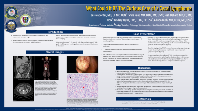
Introduction: The most common primary colorectal malignancy is adenocarcinoma. Lymphoma is an exceedingly rare diagnosis comprising only 0.2-1% of all colonic malignancies. We present the case of a 25-year-old male diagnosed with stage IV high-grade B-cell lymphoma involving a large mesenteric mass with extension to the cecum and small bowel.
Case Description/Methods: A previously healthy 25-year-old male presented to the emergency department with two months of generalized abdominal pain, nausea and vomiting, and a 15-pound weight loss. Physical exam was significant for mild tenderness of the epigastrium and left lower quadrant. Lab work with a complete blood count, hepatic function panel and right upper quadrant ultrasound were normal. Imaging with a Computed Tomography (CT) abdomen was significant for a large right-sided retroperitoneal/mesenteric mass encompassing the second portion of the duodenum and pancreas and abutting the cecum. Evaluation with a testicular ultrasound, AFP, bHCG, and CEA were normal.
An EGD showed circumferential narrowing of the distal third of the duodenum with normal appearing mucosa, concerning for extrinsic compression. On colonoscopy there was a large ulcerated non-obstructing mass in the cecum. Biopsy of the cecal mass demonstrated lymphoma and in conjunction with tissue obtained from an Interventional Radiology biopsy of a pericecal lymph node completed while awaiting hematopathology review of tissue of the cecal mass tissue, the final pathology results were consistent with a high-grade CD 10+ B-cell lymphoma. Complete staging with a PET/CT revealed one supradiaphragmatic lymph node in the right cardiophrenic space, in addition to known mesenteric lymphadenopathy. He was treated with Dose-Adjusted EPOCH-R consisting of etoposide, prednisone, vincristine, cyclophosphamide, doxorubicin, and rituximab with rapid resolution of all abdominal symptoms.
Discussion: Definitive diagnosis of mesenteric masses can be challenging and dependent on available resources. While lymphoma is a rare colonic histology, the gastrointestinal tract is the most common site of extra nodal lymphoma. Common predisposing conditions include H. pylori, EBV, immunosuppression, celiac disease, and inflammatory bowel disease. We present a case of high-grade B cell lymphoma involving the mesentery and cecum amenable to endoscopic sampling. The cecal mass tissue and subsequent molecular testing facilitated diagnosis and expeditious treatment in a young patient.

Disclosures:
Jessica Corder, MD, Shira Paul, MD, Joshua Dehart, MD, Allison Bush, MD. P1739 - What Could It B? The Curious Case of a Cecal Lymphoma, ACG 2023 Annual Scientific Meeting Abstracts. Vancouver, BC, Canada: American College of Gastroenterology.
Naval Medical Center, Portsmouth, VA
Introduction: The most common primary colorectal malignancy is adenocarcinoma. Lymphoma is an exceedingly rare diagnosis comprising only 0.2-1% of all colonic malignancies. We present the case of a 25-year-old male diagnosed with stage IV high-grade B-cell lymphoma involving a large mesenteric mass with extension to the cecum and small bowel.
Case Description/Methods: A previously healthy 25-year-old male presented to the emergency department with two months of generalized abdominal pain, nausea and vomiting, and a 15-pound weight loss. Physical exam was significant for mild tenderness of the epigastrium and left lower quadrant. Lab work with a complete blood count, hepatic function panel and right upper quadrant ultrasound were normal. Imaging with a Computed Tomography (CT) abdomen was significant for a large right-sided retroperitoneal/mesenteric mass encompassing the second portion of the duodenum and pancreas and abutting the cecum. Evaluation with a testicular ultrasound, AFP, bHCG, and CEA were normal.
An EGD showed circumferential narrowing of the distal third of the duodenum with normal appearing mucosa, concerning for extrinsic compression. On colonoscopy there was a large ulcerated non-obstructing mass in the cecum. Biopsy of the cecal mass demonstrated lymphoma and in conjunction with tissue obtained from an Interventional Radiology biopsy of a pericecal lymph node completed while awaiting hematopathology review of tissue of the cecal mass tissue, the final pathology results were consistent with a high-grade CD 10+ B-cell lymphoma. Complete staging with a PET/CT revealed one supradiaphragmatic lymph node in the right cardiophrenic space, in addition to known mesenteric lymphadenopathy. He was treated with Dose-Adjusted EPOCH-R consisting of etoposide, prednisone, vincristine, cyclophosphamide, doxorubicin, and rituximab with rapid resolution of all abdominal symptoms.
Discussion: Definitive diagnosis of mesenteric masses can be challenging and dependent on available resources. While lymphoma is a rare colonic histology, the gastrointestinal tract is the most common site of extra nodal lymphoma. Common predisposing conditions include H. pylori, EBV, immunosuppression, celiac disease, and inflammatory bowel disease. We present a case of high-grade B cell lymphoma involving the mesentery and cecum amenable to endoscopic sampling. The cecal mass tissue and subsequent molecular testing facilitated diagnosis and expeditious treatment in a young patient.

Figure: Figure 1: CT abdomen with large conglomerate soft tissue mass centered in the right lower quadrant which are is in contact with the cecum which extends to contact adjacent loops of small bowel.
Figure 2: Colonoscopy with large ulcerated non-obstructing mass in the cecum
Figure 3: CT abdomen with large soft tissue mass centered within the right lower quadrant which encases a branch of the superior mesenteric artery (arrow) without associated focal narrowing. Additional prominent lymph nodes are visualized throughout the mesentery.
Figure 4: PET/CT with large, irregularly shaped hypermetabolic nodal mass in the right lower quadrant which is in direct contact with the cecum and adjacent small bowel. Additional hypermetabolic foci scattered throughout the abdomen and pelvis and well as the right inguinofemoral region.
Disclosures:
Jessica Corder indicated no relevant financial relationships.
Shira Paul indicated no relevant financial relationships.
Joshua Dehart indicated no relevant financial relationships.
Allison Bush indicated no relevant financial relationships.
Jessica Corder, MD, Shira Paul, MD, Joshua Dehart, MD, Allison Bush, MD. P1739 - What Could It B? The Curious Case of a Cecal Lymphoma, ACG 2023 Annual Scientific Meeting Abstracts. Vancouver, BC, Canada: American College of Gastroenterology.
