Sunday Poster Session
Category: Biliary/Pancreas
P0088 - Acute Pancreatitis as the Initial Manifestation of Undiagnosed Sarcoidosis
Sunday, October 22, 2023
3:30 PM - 7:00 PM PT
Location: Exhibit Hall

Has Audio

Maria Gabriela Rubianes Guerrero, MD
Jacobi Medical Center
NY, NY
Presenting Author(s)
Award: Presidential Poster Award
Maria Gabriela Rubianes Guerrero, MD1, Felipe Ignacio Contreras-Yametti, MD2, Maria Medina Rojas, MD3, Reka Salgunan, MD1, Donald P.. Kotler, MD4, Sunny Patel, MD5
1Jacobi Medical Center, Bronx, NY; 2Wellstar Cobb Hospital, Austell, GA; 3Jacobi Medical Center, New York, NY; 4Jacobi Medical Center, Albert Einstein College of Medicine, Bronx, NY; 5Albert Einstein College of Medicine, Bronx, NY
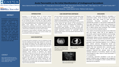
Introduction: Involvement of the pancreas in sarcoidosis is rare but has been described before, it can present as pancreatitis or mimic a pancreatic tumor. We describe a case of a woman that was admitted with pancreatitis, found to have mediastinal and hilar lymphadenopathy leading to the diagnosis of sarcoidosis.
Case Description/Methods: A 55-year-old woman presented to the ED complaining of right flank pain radiating to the right upper quadrant for a month. She also reported intermittent fever, chills, weight loss (20 pounds), daily episodes of non-bilious, and non-bloody emesis over three weeks, as well as bilateral blurry vision (worse in right eye) over 1-2 weeks.
Physical exam was notable for right upper quadrant and right costovertebral angle tenderness with mild palpation. Initial laboratory results consisted of leukopenia, and elevated transaminases, lipase was 254 U/L, ACE I was 104 U/L, bilirubin was normal. CT of chest and abdomen showed mediastinal and hilar adenopathy along with ground glass opacities in the peripheral lung fields, acute uncomplicated pancreatitis, and dilation of the pancreatic duct without evidence for acute cholecystitis. MRCP showed areas of stricture and dilation in the pancreatic duct and peripancreatic fat stranding, compatible with acute on chronic pancreatitis. EUS was notable for pancreatic parenchymal changes likely related to recent acute pancreatitis without evidence of irregular pancreatic duct or pancreatic ductal calcifications. There was no evidence of choledocholithiasis, and CBD was not dilated.
Patient received intravenous fluids and analgesics and was started on oral steroids for high suspicion of new presentation of sarcoidosis with pulmonary and extrapulmonary manifestations. She underwent endoscopic bronchial ultrasound, with transbronchial needle aspiration of 3 groups of lymph nodes. Cytology revealed lymphoid tissue with non-caseating granulomas. Despite conventional treatment for pancreatitis, patient’s symptoms didn’t improve. She only reported improvement after 4 weeks of steroids.
Discussion: Pancreatic sarcoidosis is rare and often asymptomatic; however, it can present as pancreatitis, a pancreatic mass mimicking neoplasia, or as a diffusely firm, nodular pancreas. Our patient presented with pancreatitis as the initial manifestation of sarcoidosis and her symptoms improved with steroids, which are the mainstay treatment of sarcoidosis, despite not being recommended for other causes of acute pancreatitis in current guidelines.

Disclosures:
Maria Gabriela Rubianes Guerrero, MD1, Felipe Ignacio Contreras-Yametti, MD2, Maria Medina Rojas, MD3, Reka Salgunan, MD1, Donald P.. Kotler, MD4, Sunny Patel, MD5. P0088 - Acute Pancreatitis as the Initial Manifestation of Undiagnosed Sarcoidosis, ACG 2023 Annual Scientific Meeting Abstracts. Vancouver, BC, Canada: American College of Gastroenterology.
Maria Gabriela Rubianes Guerrero, MD1, Felipe Ignacio Contreras-Yametti, MD2, Maria Medina Rojas, MD3, Reka Salgunan, MD1, Donald P.. Kotler, MD4, Sunny Patel, MD5
1Jacobi Medical Center, Bronx, NY; 2Wellstar Cobb Hospital, Austell, GA; 3Jacobi Medical Center, New York, NY; 4Jacobi Medical Center, Albert Einstein College of Medicine, Bronx, NY; 5Albert Einstein College of Medicine, Bronx, NY
Introduction: Involvement of the pancreas in sarcoidosis is rare but has been described before, it can present as pancreatitis or mimic a pancreatic tumor. We describe a case of a woman that was admitted with pancreatitis, found to have mediastinal and hilar lymphadenopathy leading to the diagnosis of sarcoidosis.
Case Description/Methods: A 55-year-old woman presented to the ED complaining of right flank pain radiating to the right upper quadrant for a month. She also reported intermittent fever, chills, weight loss (20 pounds), daily episodes of non-bilious, and non-bloody emesis over three weeks, as well as bilateral blurry vision (worse in right eye) over 1-2 weeks.
Physical exam was notable for right upper quadrant and right costovertebral angle tenderness with mild palpation. Initial laboratory results consisted of leukopenia, and elevated transaminases, lipase was 254 U/L, ACE I was 104 U/L, bilirubin was normal. CT of chest and abdomen showed mediastinal and hilar adenopathy along with ground glass opacities in the peripheral lung fields, acute uncomplicated pancreatitis, and dilation of the pancreatic duct without evidence for acute cholecystitis. MRCP showed areas of stricture and dilation in the pancreatic duct and peripancreatic fat stranding, compatible with acute on chronic pancreatitis. EUS was notable for pancreatic parenchymal changes likely related to recent acute pancreatitis without evidence of irregular pancreatic duct or pancreatic ductal calcifications. There was no evidence of choledocholithiasis, and CBD was not dilated.
Patient received intravenous fluids and analgesics and was started on oral steroids for high suspicion of new presentation of sarcoidosis with pulmonary and extrapulmonary manifestations. She underwent endoscopic bronchial ultrasound, with transbronchial needle aspiration of 3 groups of lymph nodes. Cytology revealed lymphoid tissue with non-caseating granulomas. Despite conventional treatment for pancreatitis, patient’s symptoms didn’t improve. She only reported improvement after 4 weeks of steroids.
Discussion: Pancreatic sarcoidosis is rare and often asymptomatic; however, it can present as pancreatitis, a pancreatic mass mimicking neoplasia, or as a diffusely firm, nodular pancreas. Our patient presented with pancreatitis as the initial manifestation of sarcoidosis and her symptoms improved with steroids, which are the mainstay treatment of sarcoidosis, despite not being recommended for other causes of acute pancreatitis in current guidelines.

Figure: A: CTAP with areas of stricture and dilation in the pancreatic duct. Peripancreatic fat stranding compatible with acute pancreatitis.
B: CT Chest with mediastinal and hilar adenopathy.
C: EUS showing the pancreatic body with parenchymal changes.
B: CT Chest with mediastinal and hilar adenopathy.
C: EUS showing the pancreatic body with parenchymal changes.
Disclosures:
Maria Gabriela Rubianes Guerrero indicated no relevant financial relationships.
Felipe Ignacio Contreras-Yametti indicated no relevant financial relationships.
Maria Medina Rojas indicated no relevant financial relationships.
Reka Salgunan indicated no relevant financial relationships.
Donald Kotler indicated no relevant financial relationships.
Sunny Patel indicated no relevant financial relationships.
Maria Gabriela Rubianes Guerrero, MD1, Felipe Ignacio Contreras-Yametti, MD2, Maria Medina Rojas, MD3, Reka Salgunan, MD1, Donald P.. Kotler, MD4, Sunny Patel, MD5. P0088 - Acute Pancreatitis as the Initial Manifestation of Undiagnosed Sarcoidosis, ACG 2023 Annual Scientific Meeting Abstracts. Vancouver, BC, Canada: American College of Gastroenterology.


