Sunday Poster Session
Category: Biliary/Pancreas
P0089 - Recurrent Pyogenic Cholangitis: A Case Report
Sunday, October 22, 2023
3:30 PM - 7:00 PM PT
Location: Exhibit Hall

Has Audio

Maria Gabriela Rubianes Guerrero, MD
Jacobi Medical Center
NY, NY
Presenting Author(s)
Maria Gabriela Rubianes Guerrero, MD1, Fnu Vikash, MD2, Aarushi Sudan, MD1, Sunny Patel, MD3, Donald P.. Kotler, MD2
1Jacobi Medical Center, Bronx, NY; 2Jacobi Medical Center, Albert Einstein College of Medicine, Bronx, NY; 3Albert Einstein College of Medicine, Bronx, NY
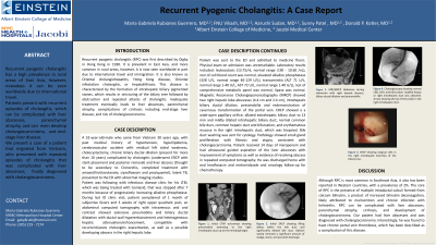
Introduction: Recurrent pyogenic cholangitis has a high prevalence in Asia but may be seen worldwide due to international travel. We present such a patient who had repeated episodes of cholangitis and liver abscesses complicated by cholangiocarcinoma.
Case Description/Methods: A 52-year-old-male who came from Vietnam 30 years ago, with a history of cholecystectomy and chronic biliary ductal dilation complicated by cholangitis, status-post ERCP with stent placement and subsequent removal, and liver abscess, presumed secondary to a liver fluke, presented to the ED with 1 month of subjective fevers and 3 weeks of right upper quadrant pain.
Physical exam was unremarkable. Laboratory results included: leukocytosis, elevated alkaline phosphatase, and elevated transaminases. An abdominal CT scan showed extensive pneumobilia and biliary ductal dilatation with ductal wall hyperenhancement compatible with recurrent/acute cholangitis exacerbation, and a possible early abscess in the right hepatic lobe. MRCP showed two right hepatic lobe abscesses, intrahepatic biliary ductal dilation, pneumobilia and redemonstration of cavernous transformation of the portal vein. ERCP demonstrated a widely opened sphincter (although patient denied prior interventions), dilated extrahepatic biliary duct to 13 mm, and erythematous mucosa in the right intrahepatic duct. A biopsy obtained via cholangioscopy of the right intrahepatic duct demonstrated adenocarcinoma. The patient received 14 days of meropenem and had aspiration of the liver abscesses with improvement of his symptoms. Subsequently, the patient was discharged home with oral antibiotics and oncology follow-up for chemotherapy.
Discussion: Although RPC is most common in Southeast Asia, it also has been reported in Western countries, with a prevalence of 2%. The core of RPC is the presence of multiple intraductal calculi formed from calcium bilirubin, a product of increased bilirubin deconjugation, likely attributed to malnutrition and chronic infection with helminths. RPC can be complicated with liver abscesses, parenchymal atrophy, cirrhosis, and development of cholangiocarcinoma. Our patient had liver abscesses and was diagnosed with cholangiocarcinoma. Interestingly, he was found to have chronic portal vein thrombosis, which has been described as a complication of this disease.

Disclosures:
Maria Gabriela Rubianes Guerrero, MD1, Fnu Vikash, MD2, Aarushi Sudan, MD1, Sunny Patel, MD3, Donald P.. Kotler, MD2. P0089 - Recurrent Pyogenic Cholangitis: A Case Report, ACG 2023 Annual Scientific Meeting Abstracts. Vancouver, BC, Canada: American College of Gastroenterology.
1Jacobi Medical Center, Bronx, NY; 2Jacobi Medical Center, Albert Einstein College of Medicine, Bronx, NY; 3Albert Einstein College of Medicine, Bronx, NY
Introduction: Recurrent pyogenic cholangitis has a high prevalence in Asia but may be seen worldwide due to international travel. We present such a patient who had repeated episodes of cholangitis and liver abscesses complicated by cholangiocarcinoma.
Case Description/Methods: A 52-year-old-male who came from Vietnam 30 years ago, with a history of cholecystectomy and chronic biliary ductal dilation complicated by cholangitis, status-post ERCP with stent placement and subsequent removal, and liver abscess, presumed secondary to a liver fluke, presented to the ED with 1 month of subjective fevers and 3 weeks of right upper quadrant pain.
Physical exam was unremarkable. Laboratory results included: leukocytosis, elevated alkaline phosphatase, and elevated transaminases. An abdominal CT scan showed extensive pneumobilia and biliary ductal dilatation with ductal wall hyperenhancement compatible with recurrent/acute cholangitis exacerbation, and a possible early abscess in the right hepatic lobe. MRCP showed two right hepatic lobe abscesses, intrahepatic biliary ductal dilation, pneumobilia and redemonstration of cavernous transformation of the portal vein. ERCP demonstrated a widely opened sphincter (although patient denied prior interventions), dilated extrahepatic biliary duct to 13 mm, and erythematous mucosa in the right intrahepatic duct. A biopsy obtained via cholangioscopy of the right intrahepatic duct demonstrated adenocarcinoma. The patient received 14 days of meropenem and had aspiration of the liver abscesses with improvement of his symptoms. Subsequently, the patient was discharged home with oral antibiotics and oncology follow-up for chemotherapy.
Discussion: Although RPC is most common in Southeast Asia, it also has been reported in Western countries, with a prevalence of 2%. The core of RPC is the presence of multiple intraductal calculi formed from calcium bilirubin, a product of increased bilirubin deconjugation, likely attributed to malnutrition and chronic infection with helminths. RPC can be complicated with liver abscesses, parenchymal atrophy, cirrhosis, and development of cholangiocarcinoma. Our patient had liver abscesses and was diagnosed with cholangiocarcinoma. Interestingly, he was found to have chronic portal vein thrombosis, which has been described as a complication of this disease.

Figure: A: Initial CTAP w/contrast showing pneumobilia extending to the right intrahepatic duct up to the hemidiaphragm.
B: Initial ERCP showing filling defect within the bile duct and significantly dilated bile duct. Balloon sweep removed a significant amount of sludge, stone, and purulent discharge.
C: MRI/MRCP Abdomen during admission with right hepatic abscess, biliary ductal dilation and pneumobilia.
D: Cholangioscopy showing normal CBD, CHD, and bifurcation. SpyBite biopsy or right intrahepatic duct was obtained. Arrow showing the tip of the probe in the right intrahepatic duct.
E: ERCP showing atypical cells in the right intrahepatic branches of the biliary tree.
Disclosures:
Maria Gabriela Rubianes Guerrero indicated no relevant financial relationships.
Fnu Vikash indicated no relevant financial relationships.
Aarushi Sudan indicated no relevant financial relationships.
Sunny Patel indicated no relevant financial relationships.
Donald Kotler indicated no relevant financial relationships.
Maria Gabriela Rubianes Guerrero, MD1, Fnu Vikash, MD2, Aarushi Sudan, MD1, Sunny Patel, MD3, Donald P.. Kotler, MD2. P0089 - Recurrent Pyogenic Cholangitis: A Case Report, ACG 2023 Annual Scientific Meeting Abstracts. Vancouver, BC, Canada: American College of Gastroenterology.
