Sunday Poster Session
Category: Biliary/Pancreas
P0095 - A Rare Case of Cystic Duct Adenocarcinoma
Sunday, October 22, 2023
3:30 PM - 7:00 PM PT
Location: Exhibit Hall

Has Audio

Misbah Safdar, MD
Bayhealth Hospital
Dover, DE
Presenting Author(s)
Misbah Safdar, MD1, Umair Chishti, MD1, Muhammad Shah Zaib, MD1, Mridul Pansari, MD1, Jing Du, MD2
1Bayhealth Hospital, Dover, DE; 2Doctors Pathology Services, Dover, DE
Introduction: Extrahepatic bile duct malignancies are about 2-3.6% of all GI malignancies. Cystic duct carcinoma is extremely rare malignancy of the biliary tract with less than 70 cases reported worldwide. (1) Adenocarcinoma is the most common, others include small cell carcinoma, carcinoid tumor, and mucin-producing carcinoma.
Case Description/Methods: 79-year-old female who was evaluated for right upper quadrant pain for several months. She underwent a HIDA scan and was found to have delayed gall bladder emptying. She underwent uneventful laparoscopic cholecystectomy. Her pathology came back positive for a 2cm adenocarcinoma arising from the cystic duct. The cystic duct margin was positive. The tumor was invading into perimuscular connective tissue on the peritoneal side and directly invaded the regional lymph node. The tumor was staged as T2A, N1, Mx. Our patient then underwent excision of the remnant cystic duct (Frozen section negative for any residual adenocarcinoma), portal lymphadenectomy and partial segment 4B and 5 liver resection. She did well post-operatively. She declined adjuvant chemotherapy. She remains under surveillance with a negative CT chest, abdomen and pelvis at 3 months follow-up.
Discussion: Cystic duct carcinoma is rare with average age of presentation 65 years (range 38-79 years). (1) Its clinical presentation is nonspecific and is usually similar to biliary calculus disease. The most common presentation is abdominal pain and advanced cystic duct carcinoma shows jaundice from the involvement of extrahepatic biliary tree. (2) The early development of symptoms with signs of gall bladder disease, slow growth and late metastasis favors a better prognosis. (1) No definite staging and diagnostic system has been established. In 1951, Farer first described diagnostic criteria to define cystic duct carcinoma. Criteria include growth restricted to the cystic duct, absence of neoplastic process in gall bladder, hepatic or CBD with histologic evidence of carcinoma. The recommended treatment is cholecystectomy with non-anatomical gall bladder fossa resection and excision of the extrahepatic bile duct along with regional lymphadenectomy. The prognosis of cystic duct carcinoma is better than other extrahepatic bile duct malignancies.
1. Lovenish Bains et al. Primary carcinoma of the cystic duct: a case report and review of classifications
2. Takenari Nakata et al. Clinical and pathological features of primary carcinoma of the cystic duct

Disclosures:
Misbah Safdar, MD1, Umair Chishti, MD1, Muhammad Shah Zaib, MD1, Mridul Pansari, MD1, Jing Du, MD2. P0095 - A Rare Case of Cystic Duct Adenocarcinoma, ACG 2023 Annual Scientific Meeting Abstracts. Vancouver, BC, Canada: American College of Gastroenterology.
1Bayhealth Hospital, Dover, DE; 2Doctors Pathology Services, Dover, DE
Introduction: Extrahepatic bile duct malignancies are about 2-3.6% of all GI malignancies. Cystic duct carcinoma is extremely rare malignancy of the biliary tract with less than 70 cases reported worldwide. (1) Adenocarcinoma is the most common, others include small cell carcinoma, carcinoid tumor, and mucin-producing carcinoma.
Case Description/Methods: 79-year-old female who was evaluated for right upper quadrant pain for several months. She underwent a HIDA scan and was found to have delayed gall bladder emptying. She underwent uneventful laparoscopic cholecystectomy. Her pathology came back positive for a 2cm adenocarcinoma arising from the cystic duct. The cystic duct margin was positive. The tumor was invading into perimuscular connective tissue on the peritoneal side and directly invaded the regional lymph node. The tumor was staged as T2A, N1, Mx. Our patient then underwent excision of the remnant cystic duct (Frozen section negative for any residual adenocarcinoma), portal lymphadenectomy and partial segment 4B and 5 liver resection. She did well post-operatively. She declined adjuvant chemotherapy. She remains under surveillance with a negative CT chest, abdomen and pelvis at 3 months follow-up.
Discussion: Cystic duct carcinoma is rare with average age of presentation 65 years (range 38-79 years). (1) Its clinical presentation is nonspecific and is usually similar to biliary calculus disease. The most common presentation is abdominal pain and advanced cystic duct carcinoma shows jaundice from the involvement of extrahepatic biliary tree. (2) The early development of symptoms with signs of gall bladder disease, slow growth and late metastasis favors a better prognosis. (1) No definite staging and diagnostic system has been established. In 1951, Farer first described diagnostic criteria to define cystic duct carcinoma. Criteria include growth restricted to the cystic duct, absence of neoplastic process in gall bladder, hepatic or CBD with histologic evidence of carcinoma. The recommended treatment is cholecystectomy with non-anatomical gall bladder fossa resection and excision of the extrahepatic bile duct along with regional lymphadenectomy. The prognosis of cystic duct carcinoma is better than other extrahepatic bile duct malignancies.
1. Lovenish Bains et al. Primary carcinoma of the cystic duct: a case report and review of classifications
2. Takenari Nakata et al. Clinical and pathological features of primary carcinoma of the cystic duct

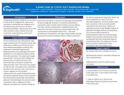
Figure: (A). Lymph node with invading carcinoma
(B).Perineural invasion -carcinoma stained red; nerve stained brown
(C&D) Residual cystic duct with invasive carcinoma
(B).Perineural invasion -carcinoma stained red; nerve stained brown
(C&D) Residual cystic duct with invasive carcinoma
Disclosures:
Misbah Safdar indicated no relevant financial relationships.
Umair Chishti indicated no relevant financial relationships.
Muhammad Shah Zaib indicated no relevant financial relationships.
Mridul Pansari indicated no relevant financial relationships.
Jing Du indicated no relevant financial relationships.
Misbah Safdar, MD1, Umair Chishti, MD1, Muhammad Shah Zaib, MD1, Mridul Pansari, MD1, Jing Du, MD2. P0095 - A Rare Case of Cystic Duct Adenocarcinoma, ACG 2023 Annual Scientific Meeting Abstracts. Vancouver, BC, Canada: American College of Gastroenterology.
