Sunday Poster Session
Category: Biliary/Pancreas
P0110 - Giant Pseudocyst Amidst: Sequential Lumen Apposing Metal Stent Placement and Chemical Necrosectomy for Giant Pseudocyst Resolution
Sunday, October 22, 2023
3:30 PM - 7:00 PM PT
Location: Exhibit Hall

Has Audio

Vikas S. Sethi, DO
Largo Medical Center
St Pete, FL
Presenting Author(s)
Vikas S. Sethi, DO1, Deep Patel, DO2, Suhail Kayyali, DO2, Yevgeniya Goltser, DO2, Bobby Jacob, MD3, Meir Mizrahi, MD2, Shivani Trivedi, DO2
1Largo Medical Center, Clearwater, FL; 2Largo Medical Center, Largo, FL; 3HCA Florida Largo Hospital, Largo, FL
Introduction: Pancreatic pseudocyst formation as a result of pancreatitis is a relatively common phenomena that has recently found new ground in treatment via endoscopic means. A concept of a giant pseudocyst is a relatively rare phenomenon and is defined as a pseudocyst with a diameter >10cm. Typically, these are addressed surgically if symptomatic and, through literature review, are sparsely addressed via endoscopic drainage. In this case, a patient presented with a complex, giant pseudocyst encasing the pancreas consisting of several smaller pseudocysts with multiple septations and internal debris. This giant pseudocyst was successfully treated with multiple lumen apposing metal stents and chemical necrosectomies in sequential fashion, avoiding the need for major surgical exploration.
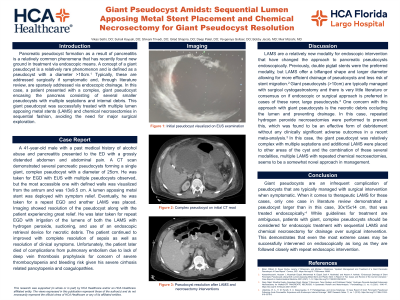
Case Description/Methods: A 41-year-old male with a past medical history of alcohol abuse and pancreatitis presented to the ED with a grossly distended abdomen and abdominal pain. A CT scan demonstrated several pancreatic pseudocysts forming a single giant, complex pseudocyst with a diameter of 25cm. He was taken for EGD with EUS with multiple pseudocysts observed, but the most accessible one with defined walls was visualized from the antrum and was 13x9.5 cm. A lumen apposing metal stent was deployed with symptom relief. Eventually, he was taken for a repeat EGD and another LAMS was placed. Imaging showed resolution of the pseudocyst along with the patient experiencing great relief. He was later taken for repeat EGD with irrigation of the lumens of both the LAMS with hydrogen peroxide, suctioning, and use of an endoscopic retrieval device for necrotic debris. Unfortunately, the patient later died of complications from pulmonary embolism due to lack of DVT prophylaxis for concern of severe thrombocytopenia and bleeding risk.
Discussion:
Giant pseudocysts are an infrequent complication of pseudocysts that are typically managed with surgical intervention when symptomatic. When it comes to therapeutic LAMS for these cases, only one case in literature review demonstrated a pseudocyst larger than in this case, 30x15x14 cm, that was treated endoscopically. While guidelines for treatment are ambiguous, patients with giant, complex pseudocysts should be considered for endoscopic treatment with sequential LAMS and chemical necrosectomy for drainage over surgical intervention. This demonstrates that even the most extreme cases may be successfully intervened on endoscopically as long as they are followed closely with repeat EGDs.

Disclosures:
Vikas S. Sethi, DO1, Deep Patel, DO2, Suhail Kayyali, DO2, Yevgeniya Goltser, DO2, Bobby Jacob, MD3, Meir Mizrahi, MD2, Shivani Trivedi, DO2. P0110 - Giant Pseudocyst Amidst: Sequential Lumen Apposing Metal Stent Placement and Chemical Necrosectomy for Giant Pseudocyst Resolution, ACG 2023 Annual Scientific Meeting Abstracts. Vancouver, BC, Canada: American College of Gastroenterology.
1Largo Medical Center, Clearwater, FL; 2Largo Medical Center, Largo, FL; 3HCA Florida Largo Hospital, Largo, FL
Introduction: Pancreatic pseudocyst formation as a result of pancreatitis is a relatively common phenomena that has recently found new ground in treatment via endoscopic means. A concept of a giant pseudocyst is a relatively rare phenomenon and is defined as a pseudocyst with a diameter >10cm. Typically, these are addressed surgically if symptomatic and, through literature review, are sparsely addressed via endoscopic drainage. In this case, a patient presented with a complex, giant pseudocyst encasing the pancreas consisting of several smaller pseudocysts with multiple septations and internal debris. This giant pseudocyst was successfully treated with multiple lumen apposing metal stents and chemical necrosectomies in sequential fashion, avoiding the need for major surgical exploration.
Case Description/Methods: A 41-year-old male with a past medical history of alcohol abuse and pancreatitis presented to the ED with a grossly distended abdomen and abdominal pain. A CT scan demonstrated several pancreatic pseudocysts forming a single giant, complex pseudocyst with a diameter of 25cm. He was taken for EGD with EUS with multiple pseudocysts observed, but the most accessible one with defined walls was visualized from the antrum and was 13x9.5 cm. A lumen apposing metal stent was deployed with symptom relief. Eventually, he was taken for a repeat EGD and another LAMS was placed. Imaging showed resolution of the pseudocyst along with the patient experiencing great relief. He was later taken for repeat EGD with irrigation of the lumens of both the LAMS with hydrogen peroxide, suctioning, and use of an endoscopic retrieval device for necrotic debris. Unfortunately, the patient later died of complications from pulmonary embolism due to lack of DVT prophylaxis for concern of severe thrombocytopenia and bleeding risk.
Discussion:
Giant pseudocysts are an infrequent complication of pseudocysts that are typically managed with surgical intervention when symptomatic. When it comes to therapeutic LAMS for these cases, only one case in literature review demonstrated a pseudocyst larger than in this case, 30x15x14 cm, that was treated endoscopically. While guidelines for treatment are ambiguous, patients with giant, complex pseudocysts should be considered for endoscopic treatment with sequential LAMS and chemical necrosectomy for drainage over surgical intervention. This demonstrates that even the most extreme cases may be successfully intervened on endoscopically as long as they are followed closely with repeat EGDs.

Figure: Image 1. Giant complex pseudocyst with a diameter of 25cm
Disclosures:
Vikas Sethi indicated no relevant financial relationships.
Deep Patel indicated no relevant financial relationships.
Suhail Kayyali indicated no relevant financial relationships.
Yevgeniya Goltser indicated no relevant financial relationships.
Bobby Jacob indicated no relevant financial relationships.
Meir Mizrahi: Boston Scientific – Consultant. COOK medical – Consultant. Fujifilm – Consultant. Medtronic – Consultant.
Shivani Trivedi indicated no relevant financial relationships.
Vikas S. Sethi, DO1, Deep Patel, DO2, Suhail Kayyali, DO2, Yevgeniya Goltser, DO2, Bobby Jacob, MD3, Meir Mizrahi, MD2, Shivani Trivedi, DO2. P0110 - Giant Pseudocyst Amidst: Sequential Lumen Apposing Metal Stent Placement and Chemical Necrosectomy for Giant Pseudocyst Resolution, ACG 2023 Annual Scientific Meeting Abstracts. Vancouver, BC, Canada: American College of Gastroenterology.
