Sunday Poster Session
Category: Biliary/Pancreas
P0128 - Isolated Brain Metastasis as Presenting Feature of Pancreatic Adenocarcinoma
Sunday, October 22, 2023
3:30 PM - 7:00 PM PT
Location: Exhibit Hall

Has Audio

Anusha Singhania, MBBS
Swedish Medical Center
Seattle, WA
Presenting Author(s)
Anusha Singhania, MBBS1, Shreya Chopra, MBBS1, Tavishi Girotra, MBBS1, Rajnish Mishra, MD1, Mohit Girotra, MD2
1Swedish Medical Center, Seattle, WA; 2Swedish Medical Center, Washington State University Elson S. Floyd School of Medicine, Seattle, WA
Introduction: Brain metastases are generally rare with GI cancers, especially with pancreatic cancer (PC) (incidence ~ 0.33–0.57%), leading to a paucity of literature on this. Our report adds to the only 3 previously reported cases of PC diagnosed after detection of brain metastases, of which 2 arose from prior intraductal papillary mucinous neoplasms (IPMN).
Case Description/Methods: A 67-year-old male, non-smoker, with a history of hypertension, polycystic kidney and liver disease, old stroke, DVT/ pulmonary embolism on apixaban, chronic renal insufficiency and chronic urinary retention was brought with altered mental status for 2 days. Accompanying family did not endorse any fever, headache, seizures, abdominal pain, unintentional loss of weight or appetite, jaundice, dark urine, nausea, or diarrhea.
Brain imaging found a left frontal 5 cm mass-like lesion with cortical/subcortical edema with effacement of frontal horn of left lateral ventricle and 5-6 mm midline shift, with areas of high attenuation (representing blood products and/or calcifications).
Cancer marker evaluation showed a high CA 19-9 (1139 IU/ml) and abdominal CT noted a poorly marginated 13x15x18 mm low-density in mid-body of pancreas (BOP) with downstream atrophy and ductal dilatation. Subsequent endoscopic ultrasound (EUS) revealed a 25 x 20 mm irregular heterogeneously hypoechoic BOP mass with multiple small peripancreatic lymph nodes. Fine needle biopsy of the mass with histopathology confirmed the diagnosis of pancreatic adenocarcinoma. After detailed discussion with neurosurgery and medical/radiation oncology, given his poor health/functional status, his prognosis was deemed poor, and the family elected palliative/hospice care.
Discussion: PC is a common, invariably fatal malignancy, with >80% cases having local or distant spread at time of diagnosis, most commonly to the liver, peritoneum or lungs. Brain metastasis is quite rare, mostly arising from lung, breast or skin primaries and indicates a universally ominous outcome. The incidence of brain metastasis with PC is rarer, with most cases found post-mortem. Our unique case reports a solitary brain metastatic lesion as the presenting feature of the underlying PC.
Urgent neurosurgical intervention has been reported to improve outcomes in such patients, with possible use of radiotherapy to control recurrences. As overall survival with PC improves, the incidence of neurological metastasis is expected to increase, warranting a watchful cognizance for this entity.

Disclosures:
Anusha Singhania, MBBS1, Shreya Chopra, MBBS1, Tavishi Girotra, MBBS1, Rajnish Mishra, MD1, Mohit Girotra, MD2. P0128 - Isolated Brain Metastasis as Presenting Feature of Pancreatic Adenocarcinoma, ACG 2023 Annual Scientific Meeting Abstracts. Vancouver, BC, Canada: American College of Gastroenterology.
1Swedish Medical Center, Seattle, WA; 2Swedish Medical Center, Washington State University Elson S. Floyd School of Medicine, Seattle, WA
Introduction: Brain metastases are generally rare with GI cancers, especially with pancreatic cancer (PC) (incidence ~ 0.33–0.57%), leading to a paucity of literature on this. Our report adds to the only 3 previously reported cases of PC diagnosed after detection of brain metastases, of which 2 arose from prior intraductal papillary mucinous neoplasms (IPMN).
Case Description/Methods: A 67-year-old male, non-smoker, with a history of hypertension, polycystic kidney and liver disease, old stroke, DVT/ pulmonary embolism on apixaban, chronic renal insufficiency and chronic urinary retention was brought with altered mental status for 2 days. Accompanying family did not endorse any fever, headache, seizures, abdominal pain, unintentional loss of weight or appetite, jaundice, dark urine, nausea, or diarrhea.
Brain imaging found a left frontal 5 cm mass-like lesion with cortical/subcortical edema with effacement of frontal horn of left lateral ventricle and 5-6 mm midline shift, with areas of high attenuation (representing blood products and/or calcifications).
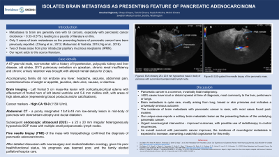
Cancer marker evaluation showed a high CA 19-9 (1139 IU/ml) and abdominal CT noted a poorly marginated 13x15x18 mm low-density in mid-body of pancreas (BOP) with downstream atrophy and ductal dilatation. Subsequent endoscopic ultrasound (EUS) revealed a 25 x 20 mm irregular heterogeneously hypoechoic BOP mass with multiple small peripancreatic lymph nodes. Fine needle biopsy of the mass with histopathology confirmed the diagnosis of pancreatic adenocarcinoma. After detailed discussion with neurosurgery and medical/radiation oncology, given his poor health/functional status, his prognosis was deemed poor, and the family elected palliative/hospice care.
Discussion: PC is a common, invariably fatal malignancy, with >80% cases having local or distant spread at time of diagnosis, most commonly to the liver, peritoneum or lungs. Brain metastasis is quite rare, mostly arising from lung, breast or skin primaries and indicates a universally ominous outcome. The incidence of brain metastasis with PC is rarer, with most cases found post-mortem. Our unique case reports a solitary brain metastatic lesion as the presenting feature of the underlying PC.
Urgent neurosurgical intervention has been reported to improve outcomes in such patients, with possible use of radiotherapy to control recurrences. As overall survival with PC improves, the incidence of neurological metastasis is expected to increase, warranting a watchful cognizance for this entity.

Figure: A. EUS showing 25 x 20.9 mm hypoechoic mass in body of pancreas with a prominent peripancreatic lymph node
B. EUS-guided fine needle biopsy of the pancreatic mass.
B. EUS-guided fine needle biopsy of the pancreatic mass.
Disclosures:
Anusha Singhania indicated no relevant financial relationships.
Shreya Chopra indicated no relevant financial relationships.
Tavishi Girotra indicated no relevant financial relationships.
Rajnish Mishra indicated no relevant financial relationships.
Mohit Girotra indicated no relevant financial relationships.
Anusha Singhania, MBBS1, Shreya Chopra, MBBS1, Tavishi Girotra, MBBS1, Rajnish Mishra, MD1, Mohit Girotra, MD2. P0128 - Isolated Brain Metastasis as Presenting Feature of Pancreatic Adenocarcinoma, ACG 2023 Annual Scientific Meeting Abstracts. Vancouver, BC, Canada: American College of Gastroenterology.
