Sunday Poster Session
Category: Biliary/Pancreas
P0131 - A Rare Case of Biliary Obstruction and Hepatic Fibrosis Secondary to Type III Choledochal Cyst
Sunday, October 22, 2023
3:30 PM - 7:00 PM PT
Location: Exhibit Hall

Has Audio

Alp S. Kahveci, MD
University of Iowa Hospital & Clinics
Iowa City, Iowa
Presenting Author(s)
Alp S. Kahveci, MD1, Darian Fard, MD2, Mohammad Abdallah, MD3, Faisal A.. Bukeirat, MD4
1University of Iowa Hospital & Clinics, Iowa City, IA; 2University of Missouri-Columbia, Columbia, MO; 3University of Missouri, Columbia, MO; 4University of Missouri at Columbia, Columbia, MO
Introduction: Choledochal cysts (CC) are congenital or acquired dilations of the biliary tree, are rather rare entities, and typically asymptomatic. They are classified into one of five forms depending on the site of dilation, most commonly type I cysts. We present a case of a type III choledochal cyst detected on endoscopic ultrasound (EUS) and endoscopic retrograde cholangiopancreatography (ERCP) in a patient with persistent post-prandial abdominal pain and markedly elevated LFTs.
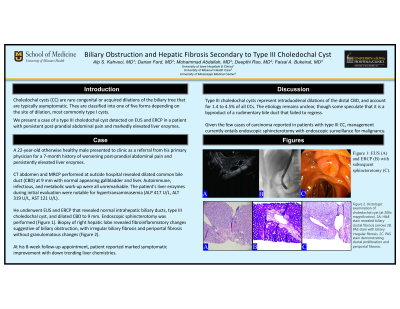
Case Description/Methods: A 22-year-old male with unremarkable past medical history presented to our gastroenterology clinic as a referral from his primary physician for a 7-month history of worsening post-prandial abdominal pain and persistently elevated liver enzymes. Computed tomography (CT) of abdomen and magnetic resonance cholangiopancreatography (MRCP) performed at outside hospital revealed dilated common bile duct (CBD) at 9 mm with normal appearing gallbladder and liver. Autoimmune, infectious, and metabolic work-up were all unremarkable. The patient's liver chemistries during initial evaluation were notable for alkaline phosphatase (ALP) 417 U/L, alanine transaminase (ALT) 319 U/L and aspartate transaminase (AST) 121 U/L. He underwent EUS and ERCP that revealed normal intrahepatic biliary ducts, type III choledochal cyst, dilated CBD to 9 mm and subsequently sphincterotomy was performed (Figure 1). Biopsy of right hepatic lobe revealed fibroinflammatory changes suggestive of biliary obstruction, with irregular biliary fibrosis and periportal fibrosis without granulomatous changes. At his 8-week follow-up appointment, patient reported marked symptomatic improvement with down trending liver chemistries.
Discussion: Type III choledochal cysts represent intraduodenal dilations of the distal CBD, and account for 1.4 to 4.5% of all CCs. The etiology remains unclear, though some speculate that it is a byproduct of a rudimentary bile duct that failed to regress. Given the limited number of cases of carcinoma reported in patients with type III CC, management currently entails endoscopic sphincterotomy with endoscopic surveillance for malignancy.

Disclosures:
Alp S. Kahveci, MD1, Darian Fard, MD2, Mohammad Abdallah, MD3, Faisal A.. Bukeirat, MD4. P0131 - A Rare Case of Biliary Obstruction and Hepatic Fibrosis Secondary to Type III Choledochal Cyst, ACG 2023 Annual Scientific Meeting Abstracts. Vancouver, BC, Canada: American College of Gastroenterology.
1University of Iowa Hospital & Clinics, Iowa City, IA; 2University of Missouri-Columbia, Columbia, MO; 3University of Missouri, Columbia, MO; 4University of Missouri at Columbia, Columbia, MO
Introduction: Choledochal cysts (CC) are congenital or acquired dilations of the biliary tree, are rather rare entities, and typically asymptomatic. They are classified into one of five forms depending on the site of dilation, most commonly type I cysts. We present a case of a type III choledochal cyst detected on endoscopic ultrasound (EUS) and endoscopic retrograde cholangiopancreatography (ERCP) in a patient with persistent post-prandial abdominal pain and markedly elevated LFTs.
Case Description/Methods: A 22-year-old male with unremarkable past medical history presented to our gastroenterology clinic as a referral from his primary physician for a 7-month history of worsening post-prandial abdominal pain and persistently elevated liver enzymes. Computed tomography (CT) of abdomen and magnetic resonance cholangiopancreatography (MRCP) performed at outside hospital revealed dilated common bile duct (CBD) at 9 mm with normal appearing gallbladder and liver. Autoimmune, infectious, and metabolic work-up were all unremarkable. The patient's liver chemistries during initial evaluation were notable for alkaline phosphatase (ALP) 417 U/L, alanine transaminase (ALT) 319 U/L and aspartate transaminase (AST) 121 U/L. He underwent EUS and ERCP that revealed normal intrahepatic biliary ducts, type III choledochal cyst, dilated CBD to 9 mm and subsequently sphincterotomy was performed (Figure 1). Biopsy of right hepatic lobe revealed fibroinflammatory changes suggestive of biliary obstruction, with irregular biliary fibrosis and periportal fibrosis without granulomatous changes. At his 8-week follow-up appointment, patient reported marked symptomatic improvement with down trending liver chemistries.
Discussion: Type III choledochal cysts represent intraduodenal dilations of the distal CBD, and account for 1.4 to 4.5% of all CCs. The etiology remains unclear, though some speculate that it is a byproduct of a rudimentary bile duct that failed to regress. Given the limited number of cases of carcinoma reported in patients with type III CC, management currently entails endoscopic sphincterotomy with endoscopic surveillance for malignancy.

Figure: Figure 1: EUS (A) and ERCP (B) with subsequent sphincterotomy (C).
Disclosures:
Alp Kahveci: Arbutus Biopharma Corp – Stock-publicly held company(excluding mutual/index funds). Bionano Genomics Inc – Stock-publicly held company(excluding mutual/index funds). Biontech SE – Stock-publicly held company(excluding mutual/index funds). Moderna Inc – Stock-publicly held company(excluding mutual/index funds). Pfizer Inc – Stock-publicly held company(excluding mutual/index funds). Senseonics Holdings Inc – Stock-publicly held company(excluding mutual/index funds).
Darian Fard indicated no relevant financial relationships.
Mohammad Abdallah indicated no relevant financial relationships.
Faisal Bukeirat indicated no relevant financial relationships.
Alp S. Kahveci, MD1, Darian Fard, MD2, Mohammad Abdallah, MD3, Faisal A.. Bukeirat, MD4. P0131 - A Rare Case of Biliary Obstruction and Hepatic Fibrosis Secondary to Type III Choledochal Cyst, ACG 2023 Annual Scientific Meeting Abstracts. Vancouver, BC, Canada: American College of Gastroenterology.
