Sunday Poster Session
Category: Biliary/Pancreas
P0139 - Assessment of Hepatic Hydrothorax: Utilizing Methylene Blue as a Diagnostic Tool
Sunday, October 22, 2023
3:30 PM - 7:00 PM PT
Location: Exhibit Hall

Has Audio

Achraf Mansour, MD
Marshall University Joan C. Edwards School of Medicine
Pasadena, CA
Presenting Author(s)
Achraf Mansour, MD1, M'hamed Turki, MD2, Abdelwahap Elghezewi, MD3, Mohammed El-Dallal, MD2, Tejas Joshi, MD3, Ahmed Sherif, MD3
1Marshall University Joan C. Edwards School of Medicine, Pasadena, CA; 2Marshall University Joan C. Edwards School of Medicine, Huntington, WV; 3Marshall University, Huntington, WV
Introduction: Pleural effusions are seen frequently in hospital setting. There are different causes for pleural effusions and the diagnostic strategy relies mainly on the suspected etiology. Hepatic hydrothorax is a relatively rare cause. To make the diagnosis of hepatic hydrothorax the presence of a peritoneo-pleural communication must be confirmed. Peritoneal scintigraphy is the method of choice but its access is limited sometimes. In this case report we discuss an alternative diagnostic tool for HH when peritoneal scintigraphy is not available.
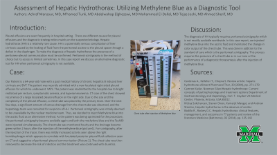
Case Description/Methods: Our Patient is a 64-year-old male with a past medical history of chronic hepatitis B induced liver cirrhosis and DVT. The patient was recently admitted with a new loculated right-sided pleural effusion for which he underwent VATS. The patient was readmitted to the hospital due to bright red blood per rectum, symptomatic anemia, and hyperammonemia. CT scan of the chest showed recurrence of a large loculated pleural effusion on the right side. Due to the size and the complexity of the pleural effusion, a chest tube was placed by the primary team. Hepatology team was consulted for suspicion of HH. Peritoneal scintigraphy was initially deemed to be unavailable by the nuclear medicine team. Thus, we decided to inject Methylene blue into the ascitic fluid as an alternative method. As the patient was being optimized for the procedure, the peritoneal scintigraphy became available again and both the methylene blue and the Tech99 were injected simultaneously. The chest tube was monitored hourly and the drainage became green within 2 hours after the injection of the methylene blue (picture1). For scintigraphy, after the injection of the tracer, there was mildly increased activity seen above the right hemidiaphragm which appears to correlate with loculated posterior pleural fluid collection seen on CT and suggestive of peritoneal-pleural communication (Picture 2). The chest tube was then removed to decrease the risk of infection.
Discussion: The diagnosis of HH typically requires peritoneal scintigraphy which is not readily available worldwide. In this case report, we injected methylene blue into the ascitic fluid and monitored the change in color output of the chest tube. This was done in addition to the standard of care which is the peritoneal scintigraphy. This process requires the presence of a chest tube as in our case or the performance of a diagnostic thoracentesis after the injection of methylene blue.

Disclosures:
Achraf Mansour, MD1, M'hamed Turki, MD2, Abdelwahap Elghezewi, MD3, Mohammed El-Dallal, MD2, Tejas Joshi, MD3, Ahmed Sherif, MD3. P0139 - Assessment of Hepatic Hydrothorax: Utilizing Methylene Blue as a Diagnostic Tool, ACG 2023 Annual Scientific Meeting Abstracts. Vancouver, BC, Canada: American College of Gastroenterology.
1Marshall University Joan C. Edwards School of Medicine, Pasadena, CA; 2Marshall University Joan C. Edwards School of Medicine, Huntington, WV; 3Marshall University, Huntington, WV
Introduction: Pleural effusions are seen frequently in hospital setting. There are different causes for pleural effusions and the diagnostic strategy relies mainly on the suspected etiology. Hepatic hydrothorax is a relatively rare cause. To make the diagnosis of hepatic hydrothorax the presence of a peritoneo-pleural communication must be confirmed. Peritoneal scintigraphy is the method of choice but its access is limited sometimes. In this case report we discuss an alternative diagnostic tool for HH when peritoneal scintigraphy is not available.
Case Description/Methods: Our Patient is a 64-year-old male with a past medical history of chronic hepatitis B induced liver cirrhosis and DVT. The patient was recently admitted with a new loculated right-sided pleural effusion for which he underwent VATS. The patient was readmitted to the hospital due to bright red blood per rectum, symptomatic anemia, and hyperammonemia. CT scan of the chest showed recurrence of a large loculated pleural effusion on the right side. Due to the size and the complexity of the pleural effusion, a chest tube was placed by the primary team. Hepatology team was consulted for suspicion of HH. Peritoneal scintigraphy was initially deemed to be unavailable by the nuclear medicine team. Thus, we decided to inject Methylene blue into the ascitic fluid as an alternative method. As the patient was being optimized for the procedure, the peritoneal scintigraphy became available again and both the methylene blue and the Tech99 were injected simultaneously. The chest tube was monitored hourly and the drainage became green within 2 hours after the injection of the methylene blue (picture1). For scintigraphy, after the injection of the tracer, there was mildly increased activity seen above the right hemidiaphragm which appears to correlate with loculated posterior pleural fluid collection seen on CT and suggestive of peritoneal-pleural communication (Picture 2). The chest tube was then removed to decrease the risk of infection.
Discussion: The diagnosis of HH typically requires peritoneal scintigraphy which is not readily available worldwide. In this case report, we injected methylene blue into the ascitic fluid and monitored the change in color output of the chest tube. This was done in addition to the standard of care which is the peritoneal scintigraphy. This process requires the presence of a chest tube as in our case or the performance of a diagnostic thoracentesis after the injection of methylene blue.

Figure: Chest tube 2 hours after injection of methylene blue
Disclosures:
Achraf Mansour indicated no relevant financial relationships.
M'hamed Turki indicated no relevant financial relationships.
Abdelwahap Elghezewi indicated no relevant financial relationships.
Mohammed El-Dallal indicated no relevant financial relationships.
Tejas Joshi indicated no relevant financial relationships.
Ahmed Sherif indicated no relevant financial relationships.
Achraf Mansour, MD1, M'hamed Turki, MD2, Abdelwahap Elghezewi, MD3, Mohammed El-Dallal, MD2, Tejas Joshi, MD3, Ahmed Sherif, MD3. P0139 - Assessment of Hepatic Hydrothorax: Utilizing Methylene Blue as a Diagnostic Tool, ACG 2023 Annual Scientific Meeting Abstracts. Vancouver, BC, Canada: American College of Gastroenterology.
