Sunday Poster Session
Category: Biliary/Pancreas
P0145 - A Rare Case of Ampullary Tubulovillous Adenoma Causing Biliary Obstruction in a Young Patient
Sunday, October 22, 2023
3:30 PM - 7:00 PM PT
Location: Exhibit Hall

Has Audio
- SE
Shalva Eliava, MD
Englewood Hospital and Medical Center
Englewood, NJ
Presenting Author(s)
Shalva Eliava, MD, Danielle Pope, MD, Natasha Rastogi, MD, Tanganyika Barnes, DO, Youran Gao, MD
Englewood Hospital and Medical Center, Englewood, NJ
Introduction: Ampullary adenomas are dysplastic lesions that arise in and around the duodenal papilla with a prevalence of 0.04% to 0.12%. They may occur in the setting of familial adenomatous polyposis (FAP) or sporadically, most commonly in patients over age 40. Ampullary adenomas are often asymptomatic and incidentally discovered on endoscopy. We present a case of an asymptomatic 34-year-old woman with incidentally-noted transaminitis found to have an ampullary tubulovillous adenoma.
Case Description/Methods: A 34-year-old female with a PMH of DM, HTN, and obesity was incidentally found to have ALP 853, AST 97, ALT 233 during an ambulatory visit. She denied a history of jaundice, abdominal pain, or weight loss at that time. The patient initially underwent laboratory testing for autoimmune and infiltrative disease and was referred for an abdominal ultrasound(US). The lab workup was nonrevealing.
Abdominal US showed dilated CBD to 1.2 cm without cholelithiasis and she was referred to Gastroenterology. MRCP was recommended but delayed due to a lack of insurance coverage. In the meantime, she developed abdominal pain.
The patient eventually presented to the emergency department with progressive, severe RUQ pain. Laboratory tests showed ALT- 458, AST- 341, ALP - 2,006. Emergent MRCP revealed a polypoid ampullary soft tissue lesion extending into the duodenum and distal CBD(measuring up to 3.3 cm), associated with moderate biliary distention. She underwent endoscopic ultrasound with a biopsy of the mass and subsequent ERCP with sphincterotomy and stent placement. Biopsy confirmed ampullary adenoma with low-grade dysplasia. Given the size and location of the lesion, surgical resection was performed instead of endoscopic resection with significant improvement in her symptoms and laboratory derangements.
Discussion: In this young patient with no family or personal history of adenomatous polyposis syndrome and without presenting symptoms, we demonstrate the need to maintain a thorough differential diagnosis when approaching the workup of liver enzyme derangements. Anchoring bias towards more common diagnoses can delay the identification of rarer conditions. Additionally, our patient experienced a delay in diagnosis due to insurance issues resulting in increased morbidity, highlighting a key systems barrier involved in pursuing elective outpatient evaluation. While her pathology was benign, there is a possibility of these lesions harboring adenocarcinoma which can significantly change treatment options and prognosis.

Disclosures:
Shalva Eliava, MD, Danielle Pope, MD, Natasha Rastogi, MD, Tanganyika Barnes, DO, Youran Gao, MD. P0145 - A Rare Case of Ampullary Tubulovillous Adenoma Causing Biliary Obstruction in a Young Patient, ACG 2023 Annual Scientific Meeting Abstracts. Vancouver, BC, Canada: American College of Gastroenterology.
Englewood Hospital and Medical Center, Englewood, NJ
Introduction: Ampullary adenomas are dysplastic lesions that arise in and around the duodenal papilla with a prevalence of 0.04% to 0.12%. They may occur in the setting of familial adenomatous polyposis (FAP) or sporadically, most commonly in patients over age 40. Ampullary adenomas are often asymptomatic and incidentally discovered on endoscopy. We present a case of an asymptomatic 34-year-old woman with incidentally-noted transaminitis found to have an ampullary tubulovillous adenoma.
Case Description/Methods: A 34-year-old female with a PMH of DM, HTN, and obesity was incidentally found to have ALP 853, AST 97, ALT 233 during an ambulatory visit. She denied a history of jaundice, abdominal pain, or weight loss at that time. The patient initially underwent laboratory testing for autoimmune and infiltrative disease and was referred for an abdominal ultrasound(US). The lab workup was nonrevealing.
Abdominal US showed dilated CBD to 1.2 cm without cholelithiasis and she was referred to Gastroenterology. MRCP was recommended but delayed due to a lack of insurance coverage. In the meantime, she developed abdominal pain.
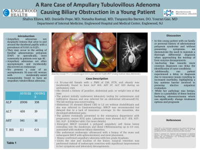
The patient eventually presented to the emergency department with progressive, severe RUQ pain. Laboratory tests showed ALT- 458, AST- 341, ALP - 2,006. Emergent MRCP revealed a polypoid ampullary soft tissue lesion extending into the duodenum and distal CBD(measuring up to 3.3 cm), associated with moderate biliary distention. She underwent endoscopic ultrasound with a biopsy of the mass and subsequent ERCP with sphincterotomy and stent placement. Biopsy confirmed ampullary adenoma with low-grade dysplasia. Given the size and location of the lesion, surgical resection was performed instead of endoscopic resection with significant improvement in her symptoms and laboratory derangements.
Discussion: In this young patient with no family or personal history of adenomatous polyposis syndrome and without presenting symptoms, we demonstrate the need to maintain a thorough differential diagnosis when approaching the workup of liver enzyme derangements. Anchoring bias towards more common diagnoses can delay the identification of rarer conditions. Additionally, our patient experienced a delay in diagnosis due to insurance issues resulting in increased morbidity, highlighting a key systems barrier involved in pursuing elective outpatient evaluation. While her pathology was benign, there is a possibility of these lesions harboring adenocarcinoma which can significantly change treatment options and prognosis.

Figure: Ampullary mass(A)
7 Fr x 7 cm double pigtail stent in place with bile flow(B)
MRCP showing moderate biliary distention(C)
7 Fr x 7 cm double pigtail stent in place with bile flow(B)
MRCP showing moderate biliary distention(C)
Disclosures:
Shalva Eliava indicated no relevant financial relationships.
Danielle Pope indicated no relevant financial relationships.
Natasha Rastogi indicated no relevant financial relationships.
Tanganyika Barnes indicated no relevant financial relationships.
Youran Gao indicated no relevant financial relationships.
Shalva Eliava, MD, Danielle Pope, MD, Natasha Rastogi, MD, Tanganyika Barnes, DO, Youran Gao, MD. P0145 - A Rare Case of Ampullary Tubulovillous Adenoma Causing Biliary Obstruction in a Young Patient, ACG 2023 Annual Scientific Meeting Abstracts. Vancouver, BC, Canada: American College of Gastroenterology.
