Sunday Poster Session
Category: Biliary/Pancreas
P0150 - Main Duct IPMN Diagnosed Using Endoscopy, EUS, and Pancreatoscopy
Sunday, October 22, 2023
3:30 PM - 7:00 PM PT
Location: Exhibit Hall

Has Audio
- DC
David L. Cheung, MD
UC Irvine
Orange, CA
Presenting Author(s)
David L. Cheung, MD1, Anastasia Chahine, MD2, Amirali Tavangar, MD2, Neil Jariwalla, MD2, Khaldoon Khirfan, MD2, Joshua Kwon, MD2, Jason Samarasena, MD2
1UC Irvine, Orange, CA; 2University of California Irvine, Orange, CA
Introduction: Intraductal papillary mucinous neoplasms (IPMNs) are pancreatic cystic neoplasms that develop within the pancreatic duct (PD). IPMNs can be present in either the main pancreatic duct or its side branches, with the risks of developing into a malignant lesion ranging from 20% to 70% in 10 years, respectively. As they are often associated with PD dilation, evidence of this on cross sectional imaging should prompt endoscopic evaluation and biopsy. One rare pathognomonic endoscopic finding for an IPMN is the “fish-mouth sign” of the ampulla of Vater. Here, we present a case of a fish mouth ampulla with positive main duct IPMN on biopsy.
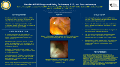
Case Description/Methods: A 72-year-old female with a history of distal pancreatectomy from a benign mass, presents for an endoscopic evaluation of a 6 mm incidental PD dilation found on CT imaging. The dilation involves both the pancreatic head and the body, but without evidence of a pancreatic mass. EGD/EUS revealed a 5.8 mm dilation within the neck and a 2.7 mm dilation within the pancreatic body, with dilation of both the duct of Wirsung and Santorini. There was also evidence of an incomplete divisum and mucinous debris within the main PD. Evaluation of the ampulla of Vater revealed a fish-mouth papilla concerning for a main duct IPMN, however, initial FNA only showed evidence of cyst debris and inflammatory cells (Image A). Given the concern for IPMN and unrevealing biopsy, the patient underwent repeat EUS and ERCP with pancreatoscopy which showed papillary structures consistent with IPMN (Image B). Repeat biopsy did show mucinous material and atypical glandular cells which was suggestive of IPMN.
Discussion: The “fish mouth” sign is a bulging, patulous Ampulla of Vater that presents with an extrusion of mucus. Seen in roughly 25% of cases of IPMN, the endoscopic finding is pathognomonic for main duct IPMN and should prompt evaluation and treatment for an IPMN. In this case, the initial biopsy was inconclusive for IPMN and there were no obvious cross sectional imaging findings that suggested presence of a mass. The presence of the “fish-mouth”, along with the presence of mucinous debris within the main PD prompted re-evaluation and biopsy with EGD/EUS. This case highlights the importance of being aware of these pathognomonic features of IPMN for diagnosis of these pre-malignant neoplasms.

Disclosures:
David L. Cheung, MD1, Anastasia Chahine, MD2, Amirali Tavangar, MD2, Neil Jariwalla, MD2, Khaldoon Khirfan, MD2, Joshua Kwon, MD2, Jason Samarasena, MD2. P0150 - Main Duct IPMN Diagnosed Using Endoscopy, EUS, and Pancreatoscopy, ACG 2023 Annual Scientific Meeting Abstracts. Vancouver, BC, Canada: American College of Gastroenterology.
1UC Irvine, Orange, CA; 2University of California Irvine, Orange, CA
Introduction: Intraductal papillary mucinous neoplasms (IPMNs) are pancreatic cystic neoplasms that develop within the pancreatic duct (PD). IPMNs can be present in either the main pancreatic duct or its side branches, with the risks of developing into a malignant lesion ranging from 20% to 70% in 10 years, respectively. As they are often associated with PD dilation, evidence of this on cross sectional imaging should prompt endoscopic evaluation and biopsy. One rare pathognomonic endoscopic finding for an IPMN is the “fish-mouth sign” of the ampulla of Vater. Here, we present a case of a fish mouth ampulla with positive main duct IPMN on biopsy.
Case Description/Methods: A 72-year-old female with a history of distal pancreatectomy from a benign mass, presents for an endoscopic evaluation of a 6 mm incidental PD dilation found on CT imaging. The dilation involves both the pancreatic head and the body, but without evidence of a pancreatic mass. EGD/EUS revealed a 5.8 mm dilation within the neck and a 2.7 mm dilation within the pancreatic body, with dilation of both the duct of Wirsung and Santorini. There was also evidence of an incomplete divisum and mucinous debris within the main PD. Evaluation of the ampulla of Vater revealed a fish-mouth papilla concerning for a main duct IPMN, however, initial FNA only showed evidence of cyst debris and inflammatory cells (Image A). Given the concern for IPMN and unrevealing biopsy, the patient underwent repeat EUS and ERCP with pancreatoscopy which showed papillary structures consistent with IPMN (Image B). Repeat biopsy did show mucinous material and atypical glandular cells which was suggestive of IPMN.
Discussion: The “fish mouth” sign is a bulging, patulous Ampulla of Vater that presents with an extrusion of mucus. Seen in roughly 25% of cases of IPMN, the endoscopic finding is pathognomonic for main duct IPMN and should prompt evaluation and treatment for an IPMN. In this case, the initial biopsy was inconclusive for IPMN and there were no obvious cross sectional imaging findings that suggested presence of a mass. The presence of the “fish-mouth”, along with the presence of mucinous debris within the main PD prompted re-evaluation and biopsy with EGD/EUS. This case highlights the importance of being aware of these pathognomonic features of IPMN for diagnosis of these pre-malignant neoplasms.

Figure: A) "Fish Mouth Sign" of Ampulla of Vater
B) Mucin and papillary structures visualized on pancreatoscopy consistent with IPMN
B) Mucin and papillary structures visualized on pancreatoscopy consistent with IPMN
Disclosures:
David Cheung indicated no relevant financial relationships.
Anastasia Chahine indicated no relevant financial relationships.
Amirali Tavangar indicated no relevant financial relationships.
Neil Jariwalla indicated no relevant financial relationships.
Khaldoon Khirfan indicated no relevant financial relationships.
Joshua Kwon indicated no relevant financial relationships.
Jason Samarasena: Applied Medical – Advisor or Review Panel Member. Boston Scientific – Consultant. Conmed – Consultant. Cook – Educational Grant. Neptune Medical – Consultant. Olympus – Consultant. Steris – Consultant.
David L. Cheung, MD1, Anastasia Chahine, MD2, Amirali Tavangar, MD2, Neil Jariwalla, MD2, Khaldoon Khirfan, MD2, Joshua Kwon, MD2, Jason Samarasena, MD2. P0150 - Main Duct IPMN Diagnosed Using Endoscopy, EUS, and Pancreatoscopy, ACG 2023 Annual Scientific Meeting Abstracts. Vancouver, BC, Canada: American College of Gastroenterology.
