Sunday Poster Session
Category: Biliary/Pancreas
P0153 - Primary Small Lymphocytic B-cell Lymphoma of Gallbladder Mimicking Acute Cholecystitis
Sunday, October 22, 2023
3:30 PM - 7:00 PM PT
Location: Exhibit Hall

Has Audio

Alaa Taha, MD
Wayne State University/Ascension Providence Rochester Hospital
Rochester, MI
Presenting Author(s)
Pranav Chalasani, MD1, Tamara Altaweel, MD2, Alaa Taha, MD3, Shahed Hussein, MD2, Laith Al-Janabi, MD4
1Wayne State University School of Medicine, Rochester Hills, MI; 2Wayne State University School of Medicine, Rochester, MI; 3Wayne State University/Ascension Providence Rochester Hospital, Rochester, MI; 4Ascension Providence Rochester Hills, Rochester, MI
Introduction: Small lymphocytic lymphoma (SLL) is a rare disease with reported incidence of less than 1% of all lymphomas. Presentation is nonspecific and can vary depending on the involved organs. Only a few cases of primary SLL involving the gallbladder are reported in literature. We present a case of a 77 year old male who presented with acute cholecystitis like symptoms, however eventually he was found to have SLL involving gallbladder and attached liver parenchyma.
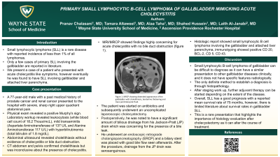
Case Description/Methods: A 77 year old male with a past medical history of prostate cancer and renal cancer presented to the hospital with severe, sharp right upper quadrant abdominal pain. Physical exam revealed a positive Murphy's sign. Laboratory workup revealed leukocytosis, mild transaminitis with hyperbilirubinemia. Abdominal ultrasound revealed cholelithiasis without evidence of cholecystitis or bile duct obstruction. CT abdomen confirmed cholelithiasis but was inconclusive regarding presence of acute cholecystitis. MRI/MRCP showed findings highly concerning for acute cholecystitis with no bile duct obstruction. The patient was started on antibiotics and subsequently underwent an uncomplicated laparoscopic cholecystectomy. Postoperatively, he was noted to have a significant amount of bilious drainage from his Jackson-Pratt (JP) drain which was concerning for the presence of a bile leak. He underwent an ERCP and a biliary stent was placed with good bile flow seen afterwards. After the procedure, drainage from the JP drain was serosanguinous. His condition improved and he was discharged the next day. Histologic report showed small lymphocytic B cell lymphoma involving the gallbladder and attached liver parenchyma, immunotyping showed positive CD 20, BCL-2, CD 5, CD 43.
Discussion: Small lymphocytic B-cell lymphoma of gallbladder can be difficult to diagnose as it can have a similar presentation to other gallbladder diseases clinically, and it does not have specific features radiologically. The only definite method to establish a diagnosis is through histopathology which is usually obtained post cholecystectomy. After staging work up, further adjuvant therapy can be started depending on the extent of the disease. Overall, SLL has a good prognosis with a reported mean survival rate of 75 months, however, there is limited literature about survival rates in gallbladder SLL. This is a rare presentation that highlights the importance of histology evaluation after cholecystectomy as it can alter the course of treatment.
Disclosures:
Pranav Chalasani, MD1, Tamara Altaweel, MD2, Alaa Taha, MD3, Shahed Hussein, MD2, Laith Al-Janabi, MD4. P0153 - Primary Small Lymphocytic B-cell Lymphoma of Gallbladder Mimicking Acute Cholecystitis, ACG 2023 Annual Scientific Meeting Abstracts. Vancouver, BC, Canada: American College of Gastroenterology.
1Wayne State University School of Medicine, Rochester Hills, MI; 2Wayne State University School of Medicine, Rochester, MI; 3Wayne State University/Ascension Providence Rochester Hospital, Rochester, MI; 4Ascension Providence Rochester Hills, Rochester, MI
Introduction: Small lymphocytic lymphoma (SLL) is a rare disease with reported incidence of less than 1% of all lymphomas. Presentation is nonspecific and can vary depending on the involved organs. Only a few cases of primary SLL involving the gallbladder are reported in literature. We present a case of a 77 year old male who presented with acute cholecystitis like symptoms, however eventually he was found to have SLL involving gallbladder and attached liver parenchyma.
Case Description/Methods: A 77 year old male with a past medical history of prostate cancer and renal cancer presented to the hospital with severe, sharp right upper quadrant abdominal pain. Physical exam revealed a positive Murphy's sign. Laboratory workup revealed leukocytosis, mild transaminitis with hyperbilirubinemia. Abdominal ultrasound revealed cholelithiasis without evidence of cholecystitis or bile duct obstruction. CT abdomen confirmed cholelithiasis but was inconclusive regarding presence of acute cholecystitis. MRI/MRCP showed findings highly concerning for acute cholecystitis with no bile duct obstruction. The patient was started on antibiotics and subsequently underwent an uncomplicated laparoscopic cholecystectomy. Postoperatively, he was noted to have a significant amount of bilious drainage from his Jackson-Pratt (JP) drain which was concerning for the presence of a bile leak. He underwent an ERCP and a biliary stent was placed with good bile flow seen afterwards. After the procedure, drainage from the JP drain was serosanguinous. His condition improved and he was discharged the next day. Histologic report showed small lymphocytic B cell lymphoma involving the gallbladder and attached liver parenchyma, immunotyping showed positive CD 20, BCL-2, CD 5, CD 43.
Discussion: Small lymphocytic B-cell lymphoma of gallbladder can be difficult to diagnose as it can have a similar presentation to other gallbladder diseases clinically, and it does not have specific features radiologically. The only definite method to establish a diagnosis is through histopathology which is usually obtained post cholecystectomy. After staging work up, further adjuvant therapy can be started depending on the extent of the disease. Overall, SLL has a good prognosis with a reported mean survival rate of 75 months, however, there is limited literature about survival rates in gallbladder SLL. This is a rare presentation that highlights the importance of histology evaluation after cholecystectomy as it can alter the course of treatment.
Disclosures:
Pranav Chalasani indicated no relevant financial relationships.
Tamara Altaweel indicated no relevant financial relationships.
Alaa Taha indicated no relevant financial relationships.
Shahed Hussein indicated no relevant financial relationships.
Laith Al-Janabi indicated no relevant financial relationships.
Pranav Chalasani, MD1, Tamara Altaweel, MD2, Alaa Taha, MD3, Shahed Hussein, MD2, Laith Al-Janabi, MD4. P0153 - Primary Small Lymphocytic B-cell Lymphoma of Gallbladder Mimicking Acute Cholecystitis, ACG 2023 Annual Scientific Meeting Abstracts. Vancouver, BC, Canada: American College of Gastroenterology.
