Sunday Poster Session
Category: Colon
P0216 - Metastatic Breast Cancer Masquerading as Colonic Mass
Sunday, October 22, 2023
3:30 PM - 7:00 PM PT
Location: Exhibit Hall

Has Audio

Zainab Mujahid, MD
University of Maryland Capital Regional Health
Lorton, VA
Presenting Author(s)
Zainab Mujahid, MD1, Omer Bajwa, MD2, Varinder Bansro, MD2, Yahia Tagouri, MD3, Gurdeep Chhabra, MD2, Homayoon Mahjoob, MD2
1University of Maryland Capital Regional Health, Lorton, VA; 2University of Maryland Capital Regional Medical Center, Largo, MD; 3MedStar Montgomery Medical Center, Olney, MD
Introduction: Breast cancer screening is a widely utilized screening test allowing for early intervention, but metastatic disease accounts for the high mortality rate. The location of metastatic presentation is variable; influenced by cell subtype and histology. Understanding cell markers and signaling pathway can assist in highlighting key pathways in metastasis and glean an understanding of these divergent behaviors.
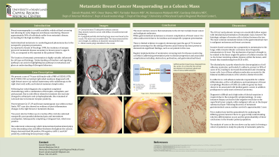
Case Description/Methods: A 74 y.o.f presents to her oncologist 2 years post-treatment of ER+, PR-, HER2/neu+ invasive lobular carcinoma (ILC) T3N1M0 of the right breast. She had undergone modified radical mastectomy involving LN 18/23+, locoregional radiotherapy 45/50 Gy in 25 fractions, and completion of carboplatin, trastuzumab, pertuzumab chemotherapy course, later switched to adriamycin, cyclophosphamide, and letrozole due to poor tolerance. Subsequent PET CT was negative for metabolically active tissue. On this visit cologuard+ stool prompted gastroenterology referral for colonoscopy. Imaging prior to procedure was significant for nonspecific thickening of the descending bowel. Left colonic and rectosigmoid biopsy revealed diffuse infiltrative cells CK7+, CK20-, ER+, and PR-, eluding to breast primary origin.
Discussion: The GI tract and pulmonic airways are essentially hollow organs with intraluminal presentation of metastatic mass; however the histologic subtype of invasive cells is distinct between the two. Invasive ductal carcinoma has a propensity to metastasize to the lungs, while ILC although uncommon more frequently metastasizes to GI tract. The mechanism of spread is understood to be related to the linear single-file pattern of cellular invasion vs. the former involving cellular division within the lumen, until breach thus transforming from DCIS to IDC. The dissimilarity is partly related to the loss of cell adhesion molecules including E-cadherin, a shared trait between certain GI cancers, leading to increased disseminated and circulating tumor cells. Hormone receptor status has also been studied to play a role, with increased receptor positivity explaining the propensity of mets to the ovary, viscera, and bone. Prior studies have laid the groundwork for analyzing the differing spread there is a gap in fully understanding why that differentiation occurs and the generalizability of these conclusions in the broader patient population. The analysis of cases as the one above can lay the groundwork in forming cohort of patients to study the polarity of metastatic patterns.

Disclosures:
Zainab Mujahid, MD1, Omer Bajwa, MD2, Varinder Bansro, MD2, Yahia Tagouri, MD3, Gurdeep Chhabra, MD2, Homayoon Mahjoob, MD2. P0216 - Metastatic Breast Cancer Masquerading as Colonic Mass, ACG 2023 Annual Scientific Meeting Abstracts. Vancouver, BC, Canada: American College of Gastroenterology.
1University of Maryland Capital Regional Health, Lorton, VA; 2University of Maryland Capital Regional Medical Center, Largo, MD; 3MedStar Montgomery Medical Center, Olney, MD
Introduction: Breast cancer screening is a widely utilized screening test allowing for early intervention, but metastatic disease accounts for the high mortality rate. The location of metastatic presentation is variable; influenced by cell subtype and histology. Understanding cell markers and signaling pathway can assist in highlighting key pathways in metastasis and glean an understanding of these divergent behaviors.
Case Description/Methods: A 74 y.o.f presents to her oncologist 2 years post-treatment of ER+, PR-, HER2/neu+ invasive lobular carcinoma (ILC) T3N1M0 of the right breast. She had undergone modified radical mastectomy involving LN 18/23+, locoregional radiotherapy 45/50 Gy in 25 fractions, and completion of carboplatin, trastuzumab, pertuzumab chemotherapy course, later switched to adriamycin, cyclophosphamide, and letrozole due to poor tolerance. Subsequent PET CT was negative for metabolically active tissue. On this visit cologuard+ stool prompted gastroenterology referral for colonoscopy. Imaging prior to procedure was significant for nonspecific thickening of the descending bowel. Left colonic and rectosigmoid biopsy revealed diffuse infiltrative cells CK7+, CK20-, ER+, and PR-, eluding to breast primary origin.
Discussion: The GI tract and pulmonic airways are essentially hollow organs with intraluminal presentation of metastatic mass; however the histologic subtype of invasive cells is distinct between the two. Invasive ductal carcinoma has a propensity to metastasize to the lungs, while ILC although uncommon more frequently metastasizes to GI tract. The mechanism of spread is understood to be related to the linear single-file pattern of cellular invasion vs. the former involving cellular division within the lumen, until breach thus transforming from DCIS to IDC. The dissimilarity is partly related to the loss of cell adhesion molecules including E-cadherin, a shared trait between certain GI cancers, leading to increased disseminated and circulating tumor cells. Hormone receptor status has also been studied to play a role, with increased receptor positivity explaining the propensity of mets to the ovary, viscera, and bone. Prior studies have laid the groundwork for analyzing the differing spread there is a gap in fully understanding why that differentiation occurs and the generalizability of these conclusions in the broader patient population. The analysis of cases as the one above can lay the groundwork in forming cohort of patients to study the polarity of metastatic patterns.

Figure: (a)diffuse wall thickening of descending colon (b) coronal view, notable for gastric abutment of transverse colon (c) tissue sample demonstrating linear single-file cells, ER +, PR-, resembling histologically primary breast lesion
Disclosures:
Zainab Mujahid indicated no relevant financial relationships.
Omer Bajwa indicated no relevant financial relationships.
Varinder Bansro indicated no relevant financial relationships.
Yahia Tagouri indicated no relevant financial relationships.
Gurdeep Chhabra indicated no relevant financial relationships.
Homayoon Mahjoob indicated no relevant financial relationships.
Zainab Mujahid, MD1, Omer Bajwa, MD2, Varinder Bansro, MD2, Yahia Tagouri, MD3, Gurdeep Chhabra, MD2, Homayoon Mahjoob, MD2. P0216 - Metastatic Breast Cancer Masquerading as Colonic Mass, ACG 2023 Annual Scientific Meeting Abstracts. Vancouver, BC, Canada: American College of Gastroenterology.

