Sunday Poster Session
Category: Colon
P0222 - Perforation of Transverse Colon by Ventriculoperitoneal Shunt
Sunday, October 22, 2023
3:30 PM - 7:00 PM PT
Location: Exhibit Hall

Has Audio

Vanessa I. Rodriguez, MD
University of South Florida
Tampa, FL
Presenting Author(s)
Award: Presidential Poster Award
Vanessa I. Rodriguez, MD1, Akash Mathavan, MD2, Akshay Mathavan, MD2, Diana N. Rodriguez, MD2, Angela Pham, MD2
1University of South Florida, Tampa, FL; 2University of Florida, Gainesville, FL
Introduction: Placement of a ventriculoperitoneal shunt (VPS) is a common neurosurgical procedure used to treat hydrocephalus via diversion of excess cerebrospinal fluid into the peritoneal cavity. Perforation of the bowel by catheter tubing of a migrated VPS is a rare complication. Although rare, it significantly increases the mortality rate. Therefore, familiarity with the clinical presentation of bowel perforation secondary to VPS placement is essential for early diagnosis and treatment.
Case Description/Methods: A man in his 40s with a history of congenital hydrocephalus and multiple VPS placements presented to the hospital for evaluation of abdominal pain. On presentation, he reported severe left-sided abdominal pain that radiated to the right side and limited mobility. Computed tomography (CT) of abdomen and pelvis showed the right VPS catheter curled in the right lower quadrant with no discontinuity or kinking. Four months prior to presentation, the patient’s right transfrontal VPS was revised. The patient was admitted due to concern of recurrent VPS-associated infection with inadequate source control. He received two weeks of empiric IV antibiotic therapy during the course of admission. Cultures showed no growth. A CT of the abdomen and pelvis 17 days after admission was notable for the right VPS entering the abdomen and extending along the region of the transverse colon (Figure 1A). However, intra-colonic shunting was deemed unlikely as the patient remained afebrile, hemodynamically stable, and exhibited no signs of peritonitis. The patient underwent colonoscopic evaluation, which revealed erosion of the VPS into the wall of the transverse colon (Figure 1B). Both the luminal and extraluminal portions were removed. The neurosurgical team externalized the remaining portion of the VPS. The patient recovered with no complications and resolution of abdominal pain.
Discussion: Rates of complication following VPS placement are considerably high. Visceral perforation secondary to shunt migration is a rare complication that poses significant elevations in hospital mortality. Patients with perforation are often asymptomatic, therefore, early diagnosis contributes to a decrease in mortality. In this case, the patient experienced persistent lower abdominal pain, but otherwise remained afebrile and hemodynamically stable. This report emphasizes the nonspecific, prolonged clinical nature of VPS-associated intestinal perforation, highlighting the need for provider awareness to ensure timely detection.

Disclosures:
Vanessa I. Rodriguez, MD1, Akash Mathavan, MD2, Akshay Mathavan, MD2, Diana N. Rodriguez, MD2, Angela Pham, MD2. P0222 - Perforation of Transverse Colon by Ventriculoperitoneal Shunt, ACG 2023 Annual Scientific Meeting Abstracts. Vancouver, BC, Canada: American College of Gastroenterology.
Vanessa I. Rodriguez, MD1, Akash Mathavan, MD2, Akshay Mathavan, MD2, Diana N. Rodriguez, MD2, Angela Pham, MD2
1University of South Florida, Tampa, FL; 2University of Florida, Gainesville, FL
Introduction: Placement of a ventriculoperitoneal shunt (VPS) is a common neurosurgical procedure used to treat hydrocephalus via diversion of excess cerebrospinal fluid into the peritoneal cavity. Perforation of the bowel by catheter tubing of a migrated VPS is a rare complication. Although rare, it significantly increases the mortality rate. Therefore, familiarity with the clinical presentation of bowel perforation secondary to VPS placement is essential for early diagnosis and treatment.
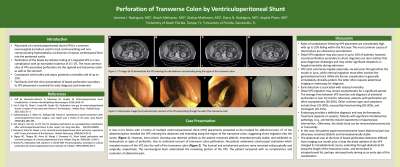
Case Description/Methods: A man in his 40s with a history of congenital hydrocephalus and multiple VPS placements presented to the hospital for evaluation of abdominal pain. On presentation, he reported severe left-sided abdominal pain that radiated to the right side and limited mobility. Computed tomography (CT) of abdomen and pelvis showed the right VPS catheter curled in the right lower quadrant with no discontinuity or kinking. Four months prior to presentation, the patient’s right transfrontal VPS was revised. The patient was admitted due to concern of recurrent VPS-associated infection with inadequate source control. He received two weeks of empiric IV antibiotic therapy during the course of admission. Cultures showed no growth. A CT of the abdomen and pelvis 17 days after admission was notable for the right VPS entering the abdomen and extending along the region of the transverse colon (Figure 1A). However, intra-colonic shunting was deemed unlikely as the patient remained afebrile, hemodynamically stable, and exhibited no signs of peritonitis. The patient underwent colonoscopic evaluation, which revealed erosion of the VPS into the wall of the transverse colon (Figure 1B). Both the luminal and extraluminal portions were removed. The neurosurgical team externalized the remaining portion of the VPS. The patient recovered with no complications and resolution of abdominal pain.
Discussion: Rates of complication following VPS placement are considerably high. Visceral perforation secondary to shunt migration is a rare complication that poses significant elevations in hospital mortality. Patients with perforation are often asymptomatic, therefore, early diagnosis contributes to a decrease in mortality. In this case, the patient experienced persistent lower abdominal pain, but otherwise remained afebrile and hemodynamically stable. This report emphasizes the nonspecific, prolonged clinical nature of VPS-associated intestinal perforation, highlighting the need for provider awareness to ensure timely detection.

Figure: Figure 1A: CT images of the abdomen demonstrating the ventriculoperitoneal shunt entering the mid-abdomen and extending along the region of the transverse colon, suggesting intra-colonic termination of the catheter.
Figure 1B: Colonoscopic images demonstrating a portion of the ventriculoperitoneal shunt penetrating through the wall of the transverse colon.
Figure 1B: Colonoscopic images demonstrating a portion of the ventriculoperitoneal shunt penetrating through the wall of the transverse colon.
Disclosures:
Vanessa Rodriguez indicated no relevant financial relationships.
Akash Mathavan indicated no relevant financial relationships.
Akshay Mathavan indicated no relevant financial relationships.
Diana Rodriguez indicated no relevant financial relationships.
Angela Pham indicated no relevant financial relationships.
Vanessa I. Rodriguez, MD1, Akash Mathavan, MD2, Akshay Mathavan, MD2, Diana N. Rodriguez, MD2, Angela Pham, MD2. P0222 - Perforation of Transverse Colon by Ventriculoperitoneal Shunt, ACG 2023 Annual Scientific Meeting Abstracts. Vancouver, BC, Canada: American College of Gastroenterology.

