Sunday Poster Session
Category: Colon
P0231 - A Rare Case of Ameboma
Sunday, October 22, 2023
3:30 PM - 7:00 PM PT
Location: Exhibit Hall

Has Audio

Heng K. Nhoung, MD
Long Island Jewish Medical Center
Forest Hills, NY
Presenting Author(s)
Heng K. Nhoung, MD, Henry K. Jen, MD
Long Island Jewish Medical Center, Forest Hills, NY
Introduction: Entamoeba histolytica is a well-known cause of infectious colitis which causes 34–50 million symptomatic infections annually worldwide. It may spread to sites such as the liver, lung, and other organs in the form of amebic abscesses, like liver abscesses. In some cases, many years after dysentery resolves, a localized infection of the colon create a segmental mass, called an “ameboma,” in patients inadequately treated during the initial course of amebic colitis.
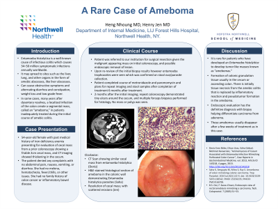
Case Description/Methods: Patient is a 54-year-old female with iron deficiency anemia presenting for evaluation of cecal mass found incidentally on prior colonoscopy showed a friable 2cm cecal mass, and CT imaging showed thickening in the cecum. The patient denied any complaints with no abdominal pain, nausea, vomiting, or diarrhea. She had no melena, hematochezia, fever/chills, or other issues. She had no family history of colon cancer or inflammatory bowel disease. She was referred to our institution for reevaluation, and possible endoscopic removal of lesion. Initially, it was unclear if this lesion was benign or inflammatory or malignant with surrounding inflammatory changes. They elected to go for repeat colonoscopy. 2 months after the initial imaging; repeat colonoscopy demonstrated tiny ulcers around the cecum, and multiple forceps biopsies performed for histology. No mass or polyp was seen. Entamoeba trophozoites were noted on biopsy and stool and ova parasites confirmed Entamoeba histolyitica. Patient completed course of metronidazole and paromomycin and plans for repeat imaging and stool samples after completion of treatment 6 months after treatment.
Discussion: It is rare for patients who have developed an Entamoeba histolyitica to develop tumor like masses known as “amebomas.” These amebomas result from the formation of colonic granulation tissue usually in the cecum or ascending colon. There is initially tissue necrosis from the amebic colitis that is replaced by inflammatory reaction and pseudotumor formation in the ameboma. They are variable in size with that can be 15cm in diameter. They can cause obstructive symptoms and alternating diarrhea and constipation, weight loss and low-grade fever. There can also be amebic liver abscess as well and it rare for them to be misdiagnosed as metastatic carcinoma of the colon. Endoscopic evaluation has the definitive diagnosis with biopsy helping differentiate carcinoma from adenoma. These amebomas usually disappear after a few weeks of treatment as in this case.
Disclosures:
Heng K. Nhoung, MD, Henry K. Jen, MD. P0231 - A Rare Case of Ameboma, ACG 2023 Annual Scientific Meeting Abstracts. Vancouver, BC, Canada: American College of Gastroenterology.
Long Island Jewish Medical Center, Forest Hills, NY
Introduction: Entamoeba histolytica is a well-known cause of infectious colitis which causes 34–50 million symptomatic infections annually worldwide. It may spread to sites such as the liver, lung, and other organs in the form of amebic abscesses, like liver abscesses. In some cases, many years after dysentery resolves, a localized infection of the colon create a segmental mass, called an “ameboma,” in patients inadequately treated during the initial course of amebic colitis.
Case Description/Methods: Patient is a 54-year-old female with iron deficiency anemia presenting for evaluation of cecal mass found incidentally on prior colonoscopy showed a friable 2cm cecal mass, and CT imaging showed thickening in the cecum. The patient denied any complaints with no abdominal pain, nausea, vomiting, or diarrhea. She had no melena, hematochezia, fever/chills, or other issues. She had no family history of colon cancer or inflammatory bowel disease. She was referred to our institution for reevaluation, and possible endoscopic removal of lesion. Initially, it was unclear if this lesion was benign or inflammatory or malignant with surrounding inflammatory changes. They elected to go for repeat colonoscopy. 2 months after the initial imaging; repeat colonoscopy demonstrated tiny ulcers around the cecum, and multiple forceps biopsies performed for histology. No mass or polyp was seen. Entamoeba trophozoites were noted on biopsy and stool and ova parasites confirmed Entamoeba histolyitica. Patient completed course of metronidazole and paromomycin and plans for repeat imaging and stool samples after completion of treatment 6 months after treatment.
Discussion: It is rare for patients who have developed an Entamoeba histolyitica to develop tumor like masses known as “amebomas.” These amebomas result from the formation of colonic granulation tissue usually in the cecum or ascending colon. There is initially tissue necrosis from the amebic colitis that is replaced by inflammatory reaction and pseudotumor formation in the ameboma. They are variable in size with that can be 15cm in diameter. They can cause obstructive symptoms and alternating diarrhea and constipation, weight loss and low-grade fever. There can also be amebic liver abscess as well and it rare for them to be misdiagnosed as metastatic carcinoma of the colon. Endoscopic evaluation has the definitive diagnosis with biopsy helping differentiate carcinoma from adenoma. These amebomas usually disappear after a few weeks of treatment as in this case.
Disclosures:
Heng Nhoung indicated no relevant financial relationships.
Henry Jen indicated no relevant financial relationships.
Heng K. Nhoung, MD, Henry K. Jen, MD. P0231 - A Rare Case of Ameboma, ACG 2023 Annual Scientific Meeting Abstracts. Vancouver, BC, Canada: American College of Gastroenterology.
