Sunday Poster Session
Category: Colon
P0238 - Lumen-apposing Metal Stent Drainage of Colonic Mucocele at Site of Anastomosis
Sunday, October 22, 2023
3:30 PM - 7:00 PM PT
Location: Exhibit Hall

Has Audio
- TG
Tyler Grantham, MD
Staten Island University Hospital
Staten Island, NY
Presenting Author(s)
Award: Presidential Poster Award
Tyler Grantham, MD1, Ahmed Elfiky, MD2, Sherif Andrawes, MD3, Jean Chalhoub, MD3, Mohammad Abureesh, MD2, Youssef El Douaihy, MD3
1Staten Island University Hospital, Staten Island, NY; 2Staten Island University Hospital Northwell Health, Staten Island, NY; 3Staten Island University Hospital, Northwell Health, Staten Island, NY
Introduction: Colonic mucoceles are an exceedingly rare occurrence and are infrequently described in literature. Appendiceal mucoceles are more commonly seen and thought to be caused by the accumulation of mucin due to appendiceal obstruction. These lesions are often asymptomatic and found incidentally, however, may present with signs of obstruction if occupying a significant portion of the lumen of the colon.
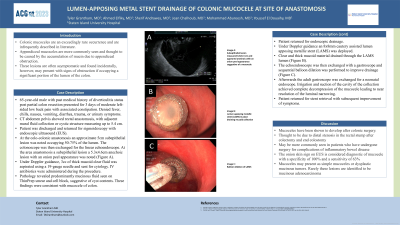
Case Description/Methods: 65-year-old male with past medical history of diverticulitis status post partial colon resection presented for 3 days of moderate left-sided low back pain with associated constipation. Denied fever, chills, nausea, vomiting, diarrhea, trauma, or urinary symptoms. CT abdomen pelvis showed rectal anastomosis, with adjacent mural fluid collection or cystic structure measuring up to 5.4 cm. Patient was discharged and returned for sigmoidoscopy with endoscopic ultrasound (EUS). At the colo-colonic anastomosis an approximate 5cm subepithelial lesion was noted occupying 50-75% of the lumen. The colonoscope was then exchanged for the linear echoendoscope. At the area anastomosis a subepithelial lesion a 5.3x4.6cm anechoic lesion with an onion peel appearance was noted (Figure A). Under Doppler guidance, 3cc of thick mucoid clear fluid was aspirated using a 19-gauge needle and sent for cytology. IV antibiotics were administered during the procedure. Pathology revealed predominantly mucinous fluid seen on ThinPrep smear and cell block, suggestive of cyst contents. These findings were consistent with mucocele of colon.
Patient returned for endoscopic drainage. Under Doppler guidance an 8x8mm cautery assisted lumen apposing metallic stent (LAMS) was deployed. Clear and thick mucoid material drained through the LAMS lumen (Figure B). The echoendosocope was then exchanged with a gastroscope and sequential balloon dilation was performed to improve drainage (Figure C). Afterwards the adult gastroscope was exchanged for a neonatal endoscope. Irrigation and suction of the cavity of the collection achieved complete decompression of the mucocele leading to near resolution of the luminal narrowing. Patient returned for stent retrieval with subsequent improvement of symptoms.
Discussion: Mucoceles have been shown to develop after colonic surgery. The onion skin sign on EUS is considered diagnostic of mucocele with a specificity of 100% and a sensitivity of 63%. Mucoceles may present as simple mucoceles or dysplastic mucinous tumors. Rarely these lesions are identified to be mucinous adenocarcinoma.

Disclosures:
Tyler Grantham, MD1, Ahmed Elfiky, MD2, Sherif Andrawes, MD3, Jean Chalhoub, MD3, Mohammad Abureesh, MD2, Youssef El Douaihy, MD3. P0238 - Lumen-apposing Metal Stent Drainage of Colonic Mucocele at Site of Anastomosis, ACG 2023 Annual Scientific Meeting Abstracts. Vancouver, BC, Canada: American College of Gastroenterology.
Tyler Grantham, MD1, Ahmed Elfiky, MD2, Sherif Andrawes, MD3, Jean Chalhoub, MD3, Mohammad Abureesh, MD2, Youssef El Douaihy, MD3
1Staten Island University Hospital, Staten Island, NY; 2Staten Island University Hospital Northwell Health, Staten Island, NY; 3Staten Island University Hospital, Northwell Health, Staten Island, NY
Introduction: Colonic mucoceles are an exceedingly rare occurrence and are infrequently described in literature. Appendiceal mucoceles are more commonly seen and thought to be caused by the accumulation of mucin due to appendiceal obstruction. These lesions are often asymptomatic and found incidentally, however, may present with signs of obstruction if occupying a significant portion of the lumen of the colon.
Case Description/Methods: 65-year-old male with past medical history of diverticulitis status post partial colon resection presented for 3 days of moderate left-sided low back pain with associated constipation. Denied fever, chills, nausea, vomiting, diarrhea, trauma, or urinary symptoms. CT abdomen pelvis showed rectal anastomosis, with adjacent mural fluid collection or cystic structure measuring up to 5.4 cm. Patient was discharged and returned for sigmoidoscopy with endoscopic ultrasound (EUS). At the colo-colonic anastomosis an approximate 5cm subepithelial lesion was noted occupying 50-75% of the lumen. The colonoscope was then exchanged for the linear echoendoscope. At the area anastomosis a subepithelial lesion a 5.3x4.6cm anechoic lesion with an onion peel appearance was noted (Figure A). Under Doppler guidance, 3cc of thick mucoid clear fluid was aspirated using a 19-gauge needle and sent for cytology. IV antibiotics were administered during the procedure. Pathology revealed predominantly mucinous fluid seen on ThinPrep smear and cell block, suggestive of cyst contents. These findings were consistent with mucocele of colon.
Patient returned for endoscopic drainage. Under Doppler guidance an 8x8mm cautery assisted lumen apposing metallic stent (LAMS) was deployed. Clear and thick mucoid material drained through the LAMS lumen (Figure B). The echoendosocope was then exchanged with a gastroscope and sequential balloon dilation was performed to improve drainage (Figure C). Afterwards the adult gastroscope was exchanged for a neonatal endoscope. Irrigation and suction of the cavity of the collection achieved complete decompression of the mucocele leading to near resolution of the luminal narrowing. Patient returned for stent retrieval with subsequent improvement of symptoms.
Discussion: Mucoceles have been shown to develop after colonic surgery. The onion skin sign on EUS is considered diagnostic of mucocele with a specificity of 100% and a sensitivity of 63%. Mucoceles may present as simple mucoceles or dysplastic mucinous tumors. Rarely these lesions are identified to be mucinous adenocarcinoma.

Figure: Figure A: Subepithelial lesion measured 6.6x5.3 cm and appeared anechoic with an onion peel appearance suggestive of a mucocele
Figure B: Lumen apposing metallic stent (LAMS) in place draining mucoid collection
Figure C: Balloon dilation of LAMS
Figure B: Lumen apposing metallic stent (LAMS) in place draining mucoid collection
Figure C: Balloon dilation of LAMS
Disclosures:
Tyler Grantham indicated no relevant financial relationships.
Ahmed Elfiky indicated no relevant financial relationships.
Sherif Andrawes indicated no relevant financial relationships.
Jean Chalhoub indicated no relevant financial relationships.
Mohammad Abureesh indicated no relevant financial relationships.
Youssef El Douaihy indicated no relevant financial relationships.
Tyler Grantham, MD1, Ahmed Elfiky, MD2, Sherif Andrawes, MD3, Jean Chalhoub, MD3, Mohammad Abureesh, MD2, Youssef El Douaihy, MD3. P0238 - Lumen-apposing Metal Stent Drainage of Colonic Mucocele at Site of Anastomosis, ACG 2023 Annual Scientific Meeting Abstracts. Vancouver, BC, Canada: American College of Gastroenterology.


